Esparto 40x serratedepi.jpg
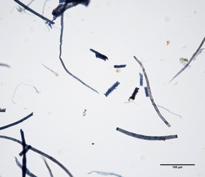
Revision as of 18:46, 18 June 2014 by JMcGlinchey (talk | contribs) (Photomicrograph showing esparto fibers and serrated epidermal cells at 400x. Sample has been stained with Graff's C-stain. Photo Credit: Jennifer McGlinchey Sexton and Paul Messier.)

Size of this preview: 692 × 599 pixels. Other resolutions: 693 × 600 pixels | 1,182 × 1,024 pixels.
Original file (1,182 × 1,024 pixels, file size: 172 KB, MIME type: image/jpeg)
Photomicrograph showing esparto fibers and serrated epidermal cells at 400x. Sample has been stained with Graff's C-stain. Photo Credit: Jennifer McGlinchey Sexton and Paul Messier.
File history
Click on a date/time to view the file as it appeared at that time.
| Date/Time | Thumbnail | Dimensions | User | Comment | |
|---|---|---|---|---|---|
| current | 18:46, 18 June 2014 | | 1,182 × 1,024 (172 KB) | JMcGlinchey (talk | contribs) | Photomicrograph showing esparto fibers and serrated epidermal cells at 400x. Sample has been stained with Graff's C-stain. Photo Credit: Jennifer McGlinchey Sexton and Paul Messier. |
You cannot overwrite this file.
File usage
The following 4 pages use this file: