Difference between revisions of "File:Rice 40x.jpg"
Jump to navigation
Jump to search
JMcGlinchey (talk | contribs) (Photomicrograph of rice pulp at 400x showing fibers, serrated epidermal cells, barrel-shaped parenchyma and other vessel elements. This sample has been stained with Graff C-Stain Photo credit: Jennifer McGlinchey Sexton and Paul Messier.) |
JMcGlinchey (talk | contribs) |
||
| Line 1: | Line 1: | ||
| − | Photomicrograph of rice pulp at 400x showing fibers, serrated epidermal cells, barrel-shaped parenchyma and other vessel elements. This sample has been stained with Graff C-Stain Photo credit: Jennifer McGlinchey Sexton and Paul Messier. | + | Photomicrograph of rice pulp at 400x showing fibers, serrated epidermal cells, barrel-shaped parenchyma and other vessel elements. This sample has been stained with [[Graff C-Stain]]. Photo credit: Jennifer McGlinchey Sexton and Paul Messier. |
Revision as of 14:17, 13 July 2015
Photomicrograph of rice pulp at 400x showing fibers, serrated epidermal cells, barrel-shaped parenchyma and other vessel elements. This sample has been stained with Graff C-Stain. Photo credit: Jennifer McGlinchey Sexton and Paul Messier.
File history
Click on a date/time to view the file as it appeared at that time.
| Date/Time | Thumbnail | Dimensions | User | Comment | |
|---|---|---|---|---|---|
| current | 10:48, 13 July 2015 | 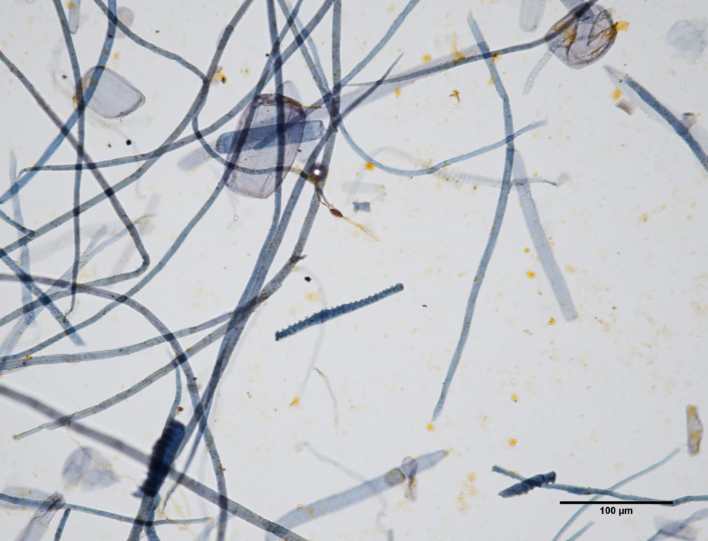 | 1,024 × 782 (99 KB) | JMcGlinchey (talk | contribs) | Photomicrograph of rice pulp at 400x showing fibers, serrated epidermal cells, barrel-shaped parenchyma and other vessel elements. This sample has been stained with Graff C-Stain Photo credit: Jennifer McGlinchey Sexton and Paul Messier. |
You cannot overwrite this file.