Difference between revisions of "X-radiography"
JLBoutaine (talk | contribs) |
m (Text replace - "== Authority ==" to "== Sources Checked for Data in Record ==") |
||
| (One intermediate revision by one other user not shown) | |||
| Line 4: | Line 4: | ||
Wilhelm Conrad Röntgen discovered the X-rays in November 1895. For this, he was distinguished by the Nobel Prize in Physics in 1901. Very shortly after his discovery, he made the first radiograph: his wife’s hand. This first image yet contained all the main possibilities of this technique: transmission through opaque media, discrimination between different materials (soft tissues, bones & metals), perceptibility of inner details of the structure. Within a month of his discovery a radiograph of a weld has already been published. Radiography became rapidly a technique used for medical diagnosis & for industrial non-destructive testing. | Wilhelm Conrad Röntgen discovered the X-rays in November 1895. For this, he was distinguished by the Nobel Prize in Physics in 1901. Very shortly after his discovery, he made the first radiograph: his wife’s hand. This first image yet contained all the main possibilities of this technique: transmission through opaque media, discrimination between different materials (soft tissues, bones & metals), perceptibility of inner details of the structure. Within a month of his discovery a radiograph of a weld has already been published. Radiography became rapidly a technique used for medical diagnosis & for industrial non-destructive testing. | ||
Radiography is one of the more used non destructive examination technique in the cltural heritage area. It is based on the transmission of X-rays through the object to be controlled. The high energy radiation is emitted by an X-ray tube powered by a hih voltage generator (from some kV to 450 kV). These radiations will penetrate different composition/density materials to varying degrees. The resulting variations of transmission of these radiations are recorded by a detector, generally a radiographic film. The resulting radiograph, owing to the differential absorption pattern, will thus permit to characterise the internal geometrical and/or compositional structure of an object, to localise & identify defects, voids, cracks, inserts, previous restorations...X-radiography is one of the major examination technique for easel paintings for examining the substrate structure (wood panel or canvas) as well as for comparing the variation in pigment types, visualising cracks, previous restorations.... | Radiography is one of the more used non destructive examination technique in the cltural heritage area. It is based on the transmission of X-rays through the object to be controlled. The high energy radiation is emitted by an X-ray tube powered by a hih voltage generator (from some kV to 450 kV). These radiations will penetrate different composition/density materials to varying degrees. The resulting variations of transmission of these radiations are recorded by a detector, generally a radiographic film. The resulting radiograph, owing to the differential absorption pattern, will thus permit to characterise the internal geometrical and/or compositional structure of an object, to localise & identify defects, voids, cracks, inserts, previous restorations...X-radiography is one of the major examination technique for easel paintings for examining the substrate structure (wood panel or canvas) as well as for comparing the variation in pigment types, visualising cracks, previous restorations.... | ||
| − | When the penetration power of the X-rays become insufficient, one can use gamma radiography or radiography with an accelerator (for instance for large stone of metal statues). | + | When the penetration power of the X-rays become insufficient, one can use [[gamma radiography]] or radiography with an accelerator (for instance for large stone of metal statues). |
== Synonyms and Related Terms == | == Synonyms and Related Terms == | ||
| Line 18: | Line 18: | ||
| − | == | + | == Sources Checked for Data in Record == |
* P.A. Ruault, Radiologie industrielle, 2 vol., Paris, Institute de Soudure (1991) | * P.A. Ruault, Radiologie industrielle, 2 vol., Paris, Institute de Soudure (1991) | ||
Revision as of 23:13, 1 May 2016

Description
Wilhelm Conrad Röntgen discovered the X-rays in November 1895. For this, he was distinguished by the Nobel Prize in Physics in 1901. Very shortly after his discovery, he made the first radiograph: his wife’s hand. This first image yet contained all the main possibilities of this technique: transmission through opaque media, discrimination between different materials (soft tissues, bones & metals), perceptibility of inner details of the structure. Within a month of his discovery a radiograph of a weld has already been published. Radiography became rapidly a technique used for medical diagnosis & for industrial non-destructive testing. Radiography is one of the more used non destructive examination technique in the cltural heritage area. It is based on the transmission of X-rays through the object to be controlled. The high energy radiation is emitted by an X-ray tube powered by a hih voltage generator (from some kV to 450 kV). These radiations will penetrate different composition/density materials to varying degrees. The resulting variations of transmission of these radiations are recorded by a detector, generally a radiographic film. The resulting radiograph, owing to the differential absorption pattern, will thus permit to characterise the internal geometrical and/or compositional structure of an object, to localise & identify defects, voids, cracks, inserts, previous restorations...X-radiography is one of the major examination technique for easel paintings for examining the substrate structure (wood panel or canvas) as well as for comparing the variation in pigment types, visualising cracks, previous restorations.... When the penetration power of the X-rays become insufficient, one can use Gamma radiography or radiography with an accelerator (for instance for large stone of metal statues).
Synonyms and Related Terms
radiographie X (Fr.); Röntgendurchstrahlung (Deut.); radiografia de raios X (Port.)
Additional Images
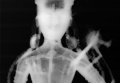
X-ray image of a statue
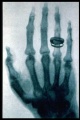
X-ray image by Roentgen


Sources Checked for Data in Record
- P.A. Ruault, Radiologie industrielle, 2 vol., Paris, Institute de Soudure (1991)
- R. Halmshaw, Industrial radiology: theory and practice (2nd edition), Chapman & Hall, London (1995)
- R.H.Bossi, F. Iddings, G.C. Wheeler, P.O. Moore, Nondestructive testing handbook, vol 4, Radiographic testing, 3rd edition, Columbus OH, ASNT, (2002)
- Anonymous Eastman-Kodak, Radiography in modern industry (4th edition), Rochester, (1980),
http://www.kodak.com/eknec/documents/87/0900688a802b3c87/Radiography-in-Modern-Industry.pdf & http://carestream.com/ndt-resources.html
- Anonymous – General Electric Inspection Technologies, Industrial radiography – Image forming techniques,
- Anonymous– Fujifilm, The fundamental of industrial radiography, http://www.fujifilm.com/products/ndt/pdf/ix-film_fundamentals_of_industrial_radiography.pdf
- M. Hours, La radiographie des oeuvres d'art, in Idées et Découvertes, Encyclopaedia Universalis, Paris, 240-243 (1978)
- M. Hours, Les secrets des chefs d'oeuvre, R. Laffont, Paris (1964)
- A. Middleton, J. Lang, Radiography of cultural material, 2nd edition, London, Butterworth – Heinemann (2005)
- A. Gilardoni, L. Ascani Orsini, R. Ascani Orsini, M. Taccani Gilardoni & al., X-rays in art - I raggi X nell'arte (2nd edition), Gilardoni, Mandello Lario (1984)
- D. Graham, T. Eddie, X-ray techniques in art galleries and museums, A. Hilger, Bristol (1985)
- E. Ravaud, La radiographie des peintures. Apport en histoire des techniques picturales & en conservation restauration, Thesis, Université Paris I Panthéon Sorbonne, Paris (February 2011)