Difference between revisions of "File:1 01 1 GypsumAlabaster STEMI 1000um.jpg"
Jump to navigation
Jump to search
| Line 1: | Line 1: | ||
| − | Photomicrograph with 1000µm measurement bar(approximate to 50x magnification). | + | Photomicrograph with 1000µm measurement bar (approximate to 50x magnification). |
Image captured using Zeiss STEMI SV 11 stereomicroscope. Pigment sample was applied to carbon tape and mounted onto aluminum examination stub. Image illuminated by 3200K raking and direct light. Image is compressed from an original 5.6 MB TIF file. | Image captured using Zeiss STEMI SV 11 stereomicroscope. Pigment sample was applied to carbon tape and mounted onto aluminum examination stub. Image illuminated by 3200K raking and direct light. Image is compressed from an original 5.6 MB TIF file. | ||
Latest revision as of 11:07, 3 January 2017
Photomicrograph with 1000µm measurement bar (approximate to 50x magnification). Image captured using Zeiss STEMI SV 11 stereomicroscope. Pigment sample was applied to carbon tape and mounted onto aluminum examination stub. Image illuminated by 3200K raking and direct light. Image is compressed from an original 5.6 MB TIF file.
File history
Click on a date/time to view the file as it appeared at that time.
| Date/Time | Thumbnail | Dimensions | User | Comment | |
|---|---|---|---|---|---|
| current | 15:08, 29 December 2016 | 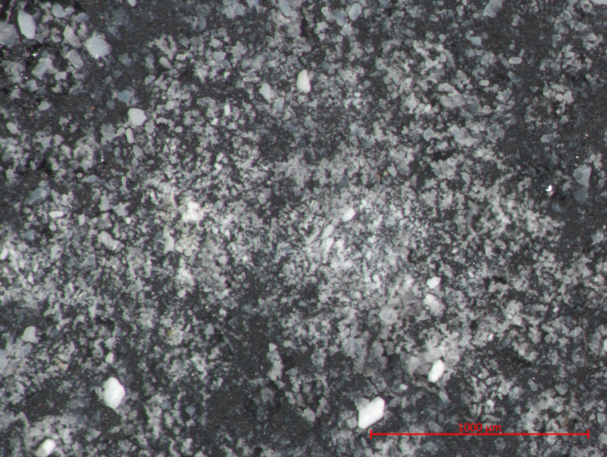 | 1,936 × 1,460 (878 KB) | MKullman (talk | contribs) | Photomicrograph with 1000µm measurement bar(approximate to 50x magnification). Image captured using Zeiss STEMI SV 11 stereomicroscope. Pigment sample was applied to carbon tape and mounted onto aluminum examination stub. Image was captured illuminat... |
You cannot overwrite this file.
File usage
The following page uses this file: