Difference between revisions of "File:1 01 9 BoneAsh SEM 100um.jpg"
Jump to navigation
Jump to search
(SEM image with 100µm measurement bar (approximate to 500x magnification) (LC) Imaged with FEI Quanta 600 scanning electron microscope (SEM) and xT microscope Server user interface. Pigment sample was applied to carbon tape and mounted onto aluminum e...) |
|||
| Line 1: | Line 1: | ||
SEM image with 100µm measurement bar (approximate to 500x magnification) (LC) | SEM image with 100µm measurement bar (approximate to 500x magnification) (LC) | ||
| − | Imaged with FEI Quanta 600 scanning electron microscope (SEM) and xT microscope Server user interface. Pigment sample was applied to carbon tape and mounted onto aluminum examination stub. Imaging was performed in a low vacuum (10 Pascal) environment and signal was collected using an FEI solid-state backscattered electron detector (BSED). Accelerating voltage: 21.4 kV | + | Imaged with FEI Quanta 600 scanning electron microscope (SEM) and xT microscope Server user interface. Pigment sample was applied to carbon tape and mounted onto aluminum examination stub. Imaging was performed in a low vacuum (10 Pascal) environment and signal was collected using an FEI solid-state backscattered electron detector (BSED). Accelerating voltage: 21.4 kV. |
Image is compressed from an original 954 KB TIF file. | Image is compressed from an original 954 KB TIF file. | ||
Latest revision as of 10:09, 10 January 2017
SEM image with 100µm measurement bar (approximate to 500x magnification) (LC) Imaged with FEI Quanta 600 scanning electron microscope (SEM) and xT microscope Server user interface. Pigment sample was applied to carbon tape and mounted onto aluminum examination stub. Imaging was performed in a low vacuum (10 Pascal) environment and signal was collected using an FEI solid-state backscattered electron detector (BSED). Accelerating voltage: 21.4 kV. Image is compressed from an original 954 KB TIF file.
File history
Click on a date/time to view the file as it appeared at that time.
| Date/Time | Thumbnail | Dimensions | User | Comment | |
|---|---|---|---|---|---|
| current | 13:44, 4 January 2017 | 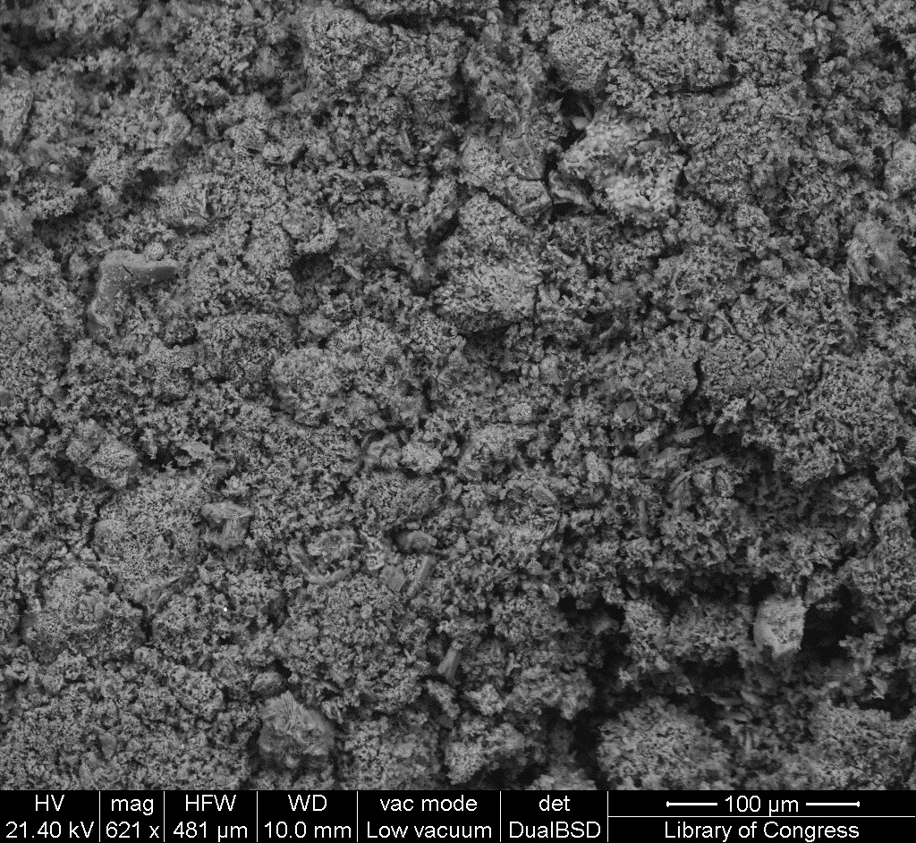 | 1,024 × 943 (566 KB) | MKullman (talk | contribs) | SEM image with 100µm measurement bar (approximate to 500x magnification) (LC) Imaged with FEI Quanta 600 scanning electron microscope (SEM) and xT microscope Server user interface. Pigment sample was applied to carbon tape and mounted onto aluminum e... |
You cannot overwrite this file.
File usage
The following page uses this file: