Difference between revisions of "Optical microscopy"
m (MDerrick moved page Optical microscope to Optical microscopy without leaving a redirect) |
|||
| Line 4: | Line 4: | ||
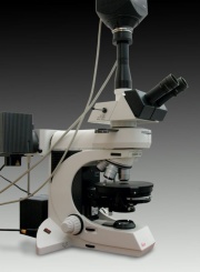
Optical microscopy (also called light microscopy) is a technique for the visual characterization of materials. It is possible to evaluate both physical and chemical properties of materials based on light interactions with a sample while observing it at magnification. Various reagents, optical filters, and preparation methods are available that may either enhance or provide additional visual cues, facilitating characterization. The basic configuration consists of two lens systems, in which the objective lens forms a magnified image of the object, which is further magnified by the eyepieces. Transparent samples are typically viewed with transmitted light, while opaque samples are observed with reflected light. Both lighting configurations may be necessary to characterize a sample. | Optical microscopy (also called light microscopy) is a technique for the visual characterization of materials. It is possible to evaluate both physical and chemical properties of materials based on light interactions with a sample while observing it at magnification. Various reagents, optical filters, and preparation methods are available that may either enhance or provide additional visual cues, facilitating characterization. The basic configuration consists of two lens systems, in which the objective lens forms a magnified image of the object, which is further magnified by the eyepieces. Transparent samples are typically viewed with transmitted light, while opaque samples are observed with reflected light. Both lighting configurations may be necessary to characterize a sample. | ||
| − | Broad ranges of artists' materials lend themselves to light microscopic analysis. Materials such as pigments, textile fibers, paper fibers, woods, and corrosion products are examined using high-magnification techniques that require a prepared sample. Information about gross surface features can be gleaned using low-magnification microscopes and do not require sampling the object. In the case of multilayered objects (for example an easel painting or polychrome sculpture), samples prepared as cross sections allow the analyst to address stratigraphy issues and questions about artist's technique. A cross section is an excised sample that has been mounted in a resin block, which is cut, ground, and polished perpendicular to the layering. The cross section is then viewed in reflected light. Details of the number of layers and their ordering, thickness, condition, and composition can be assessed. The analysis of cross sections is one of the more widely used optical microscopy techniques in conservation | + | Broad ranges of artists' materials lend themselves to light microscopic analysis. Materials such as pigments, textile fibers, paper fibers, woods, and corrosion products are examined using high-magnification techniques that require a prepared sample. Information about gross surface features can be gleaned using low-magnification microscopes and do not require sampling the object. In the case of multilayered objects (for example an easel painting or polychrome sculpture), samples prepared as cross sections allow the analyst to address stratigraphy issues and questions about artist's technique. A cross section is an excised sample that has been mounted in a resin block, which is cut, ground, and polished perpendicular to the layering. The cross section is then viewed in reflected light. Details of the number of layers and their ordering, thickness, condition, and composition can be assessed. The analysis of cross sections is one of the more widely used optical microscopy techniques in conservation. |
| + | |||
| + | The description above obtained from * National Gallery of Art, Scientific Research: [https://www.nga.gov/conservation/glossary.html Glossary of Conservation Terminology] | ||
== Synonyms and related terms == | == Synonyms and related terms == | ||
| Line 11: | Line 13: | ||
==Resources and Citations== | ==Resources and Citations== | ||
| − | + | * Wikipedia: [https://en.wikipedia.org/wiki/Optical_microscope Optical microscope] | |
* Walter C. McCrone, "Polarized Light Microscopy in Conservation: A Personal Perspective" ''JAIC'' Vol 33(2):101-14, 1994. | * Walter C. McCrone, "Polarized Light Microscopy in Conservation: A Personal Perspective" ''JAIC'' Vol 33(2):101-14, 1994. | ||
* Olympus Microscopy Resource Center at http://www.olympusmicro.com | * Olympus Microscopy Resource Center at http://www.olympusmicro.com | ||
* ASTM, Standard Terminology of Microscopy, Annual Book of ASTM Standards, Section 14, General Methods and Instrumentation, ASTM, E175, 75-78, May 1982 | * ASTM, Standard Terminology of Microscopy, Annual Book of ASTM Standards, Section 14, General Methods and Instrumentation, ASTM, E175, 75-78, May 1982 | ||
| − | + | ||
Revision as of 11:31, 20 June 2023
Description
Optical microscopy (also called light microscopy) is a technique for the visual characterization of materials. It is possible to evaluate both physical and chemical properties of materials based on light interactions with a sample while observing it at magnification. Various reagents, optical filters, and preparation methods are available that may either enhance or provide additional visual cues, facilitating characterization. The basic configuration consists of two lens systems, in which the objective lens forms a magnified image of the object, which is further magnified by the eyepieces. Transparent samples are typically viewed with transmitted light, while opaque samples are observed with reflected light. Both lighting configurations may be necessary to characterize a sample.
Broad ranges of artists' materials lend themselves to light microscopic analysis. Materials such as pigments, textile fibers, paper fibers, woods, and corrosion products are examined using high-magnification techniques that require a prepared sample. Information about gross surface features can be gleaned using low-magnification microscopes and do not require sampling the object. In the case of multilayered objects (for example an easel painting or polychrome sculpture), samples prepared as cross sections allow the analyst to address stratigraphy issues and questions about artist's technique. A cross section is an excised sample that has been mounted in a resin block, which is cut, ground, and polished perpendicular to the layering. The cross section is then viewed in reflected light. Details of the number of layers and their ordering, thickness, condition, and composition can be assessed. The analysis of cross sections is one of the more widely used optical microscopy techniques in conservation.
The description above obtained from * National Gallery of Art, Scientific Research: Glossary of Conservation Terminology
microscope optique (Fr.)
Resources and Citations
- Wikipedia: Optical microscope
- Walter C. McCrone, "Polarized Light Microscopy in Conservation: A Personal Perspective" JAIC Vol 33(2):101-14, 1994.
- Olympus Microscopy Resource Center at http://www.olympusmicro.com
- ASTM, Standard Terminology of Microscopy, Annual Book of ASTM Standards, Section 14, General Methods and Instrumentation, ASTM, E175, 75-78, May 1982
