Uploads by MKullman
Jump to navigation
Jump to search
This special page shows all uploaded files.
| Date | Name | Thumbnail | Size | Description | Versions |
|---|---|---|---|---|---|
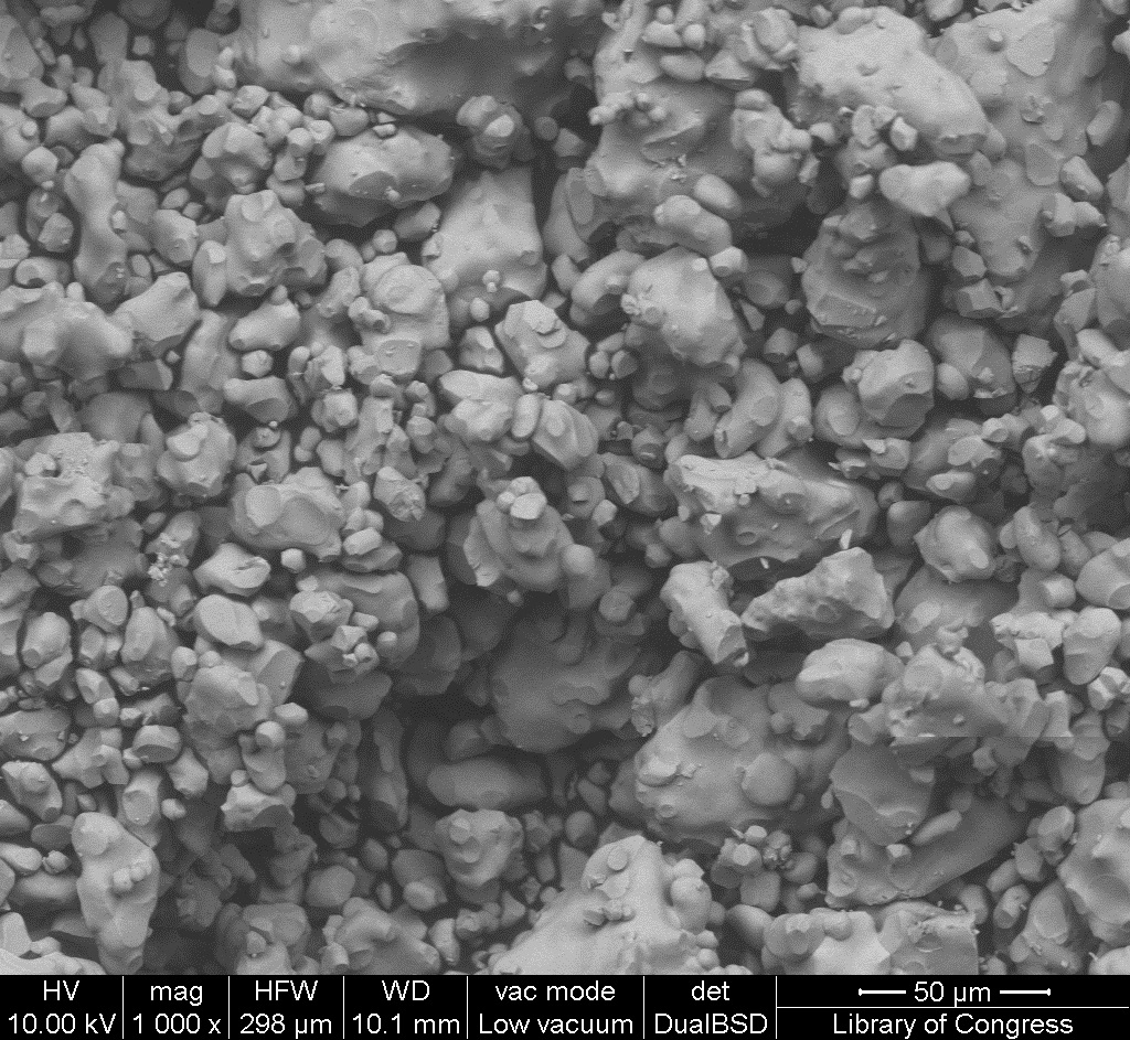
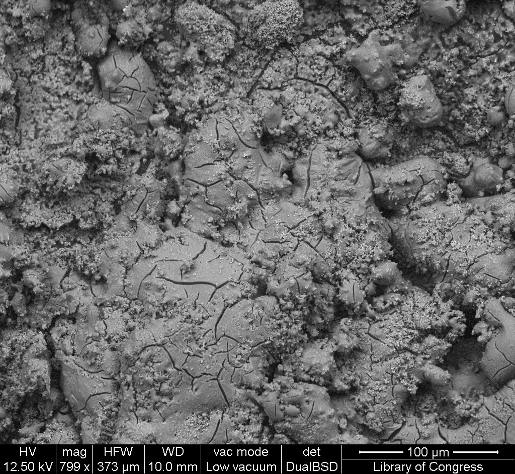
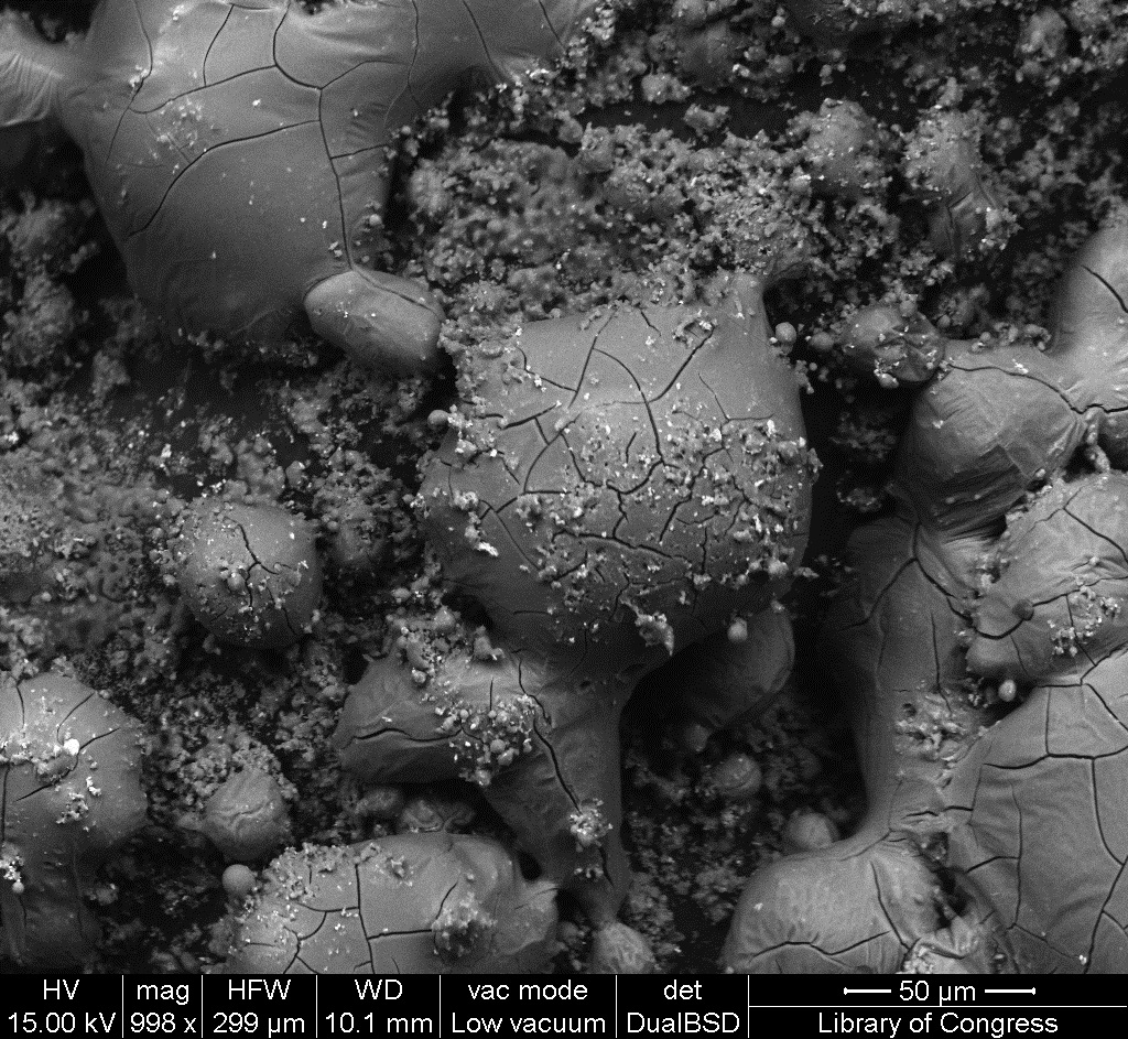
| 13:51, 12 January 2017 | 1 10 2 WhiteSpain SEM 50um.jpg (file) |  |
445 KB | SEM image with 50µm measurement bar (approximate to 1000x magnification) (LC) Imaged with FEI Quanta 600 scanning electron microscope (SEM) and xT microscope Server user interface. Pigment sample was applied to carbon tape and mounted onto aluminum e... | 1 |
| 13:50, 12 January 2017 | 1 10 2 WhiteSpain SEM 100um.jpg (file) |  |
497 KB | SEM image with 100µm measurement bar (approximate to 500x magnification) (LC) Imaged with FEI Quanta 600 scanning electron microscope (SEM) and xT microscope Server user interface. Pigment sample was applied to carbon tape and mounted onto aluminum e... | 1 |
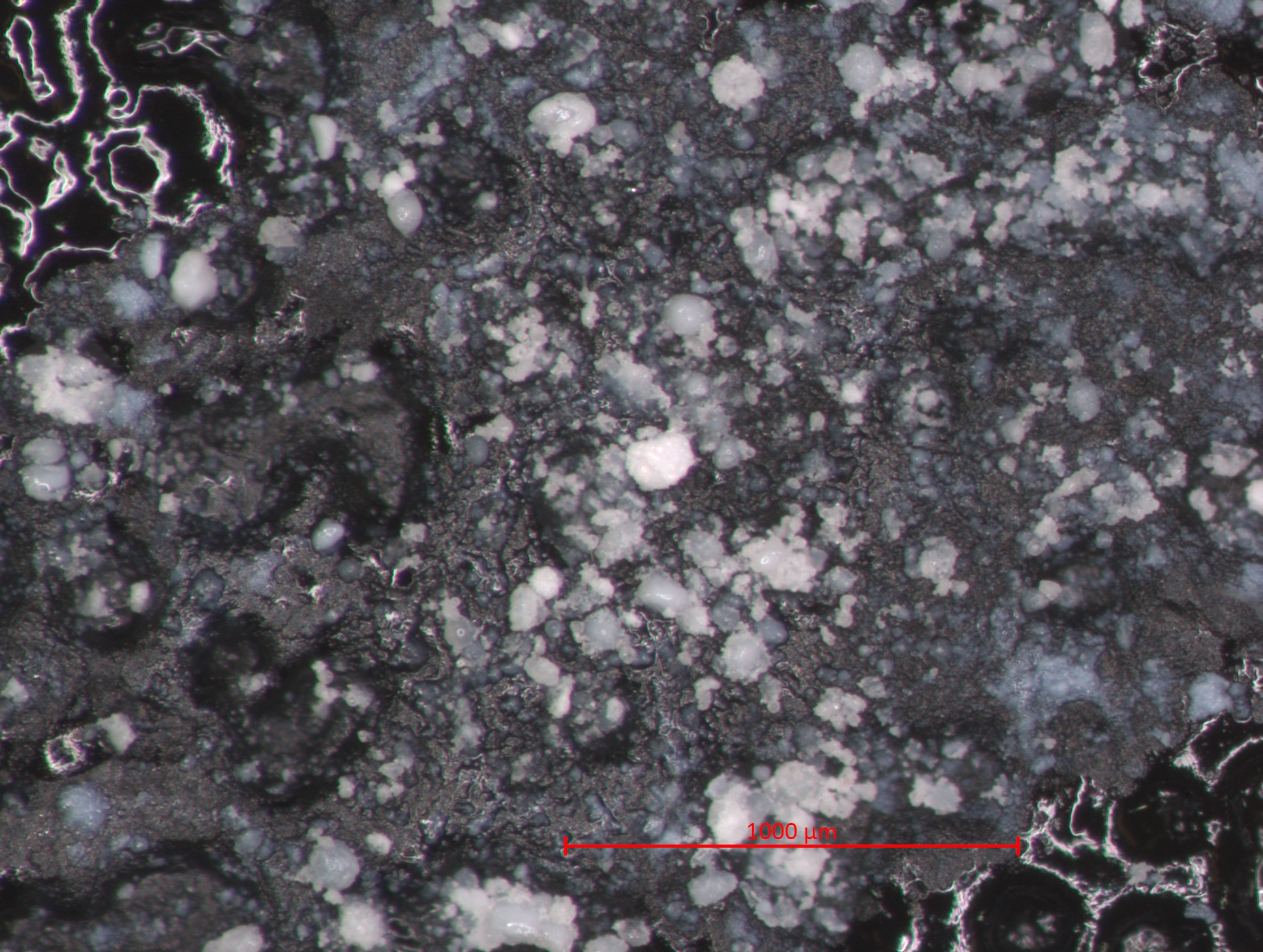
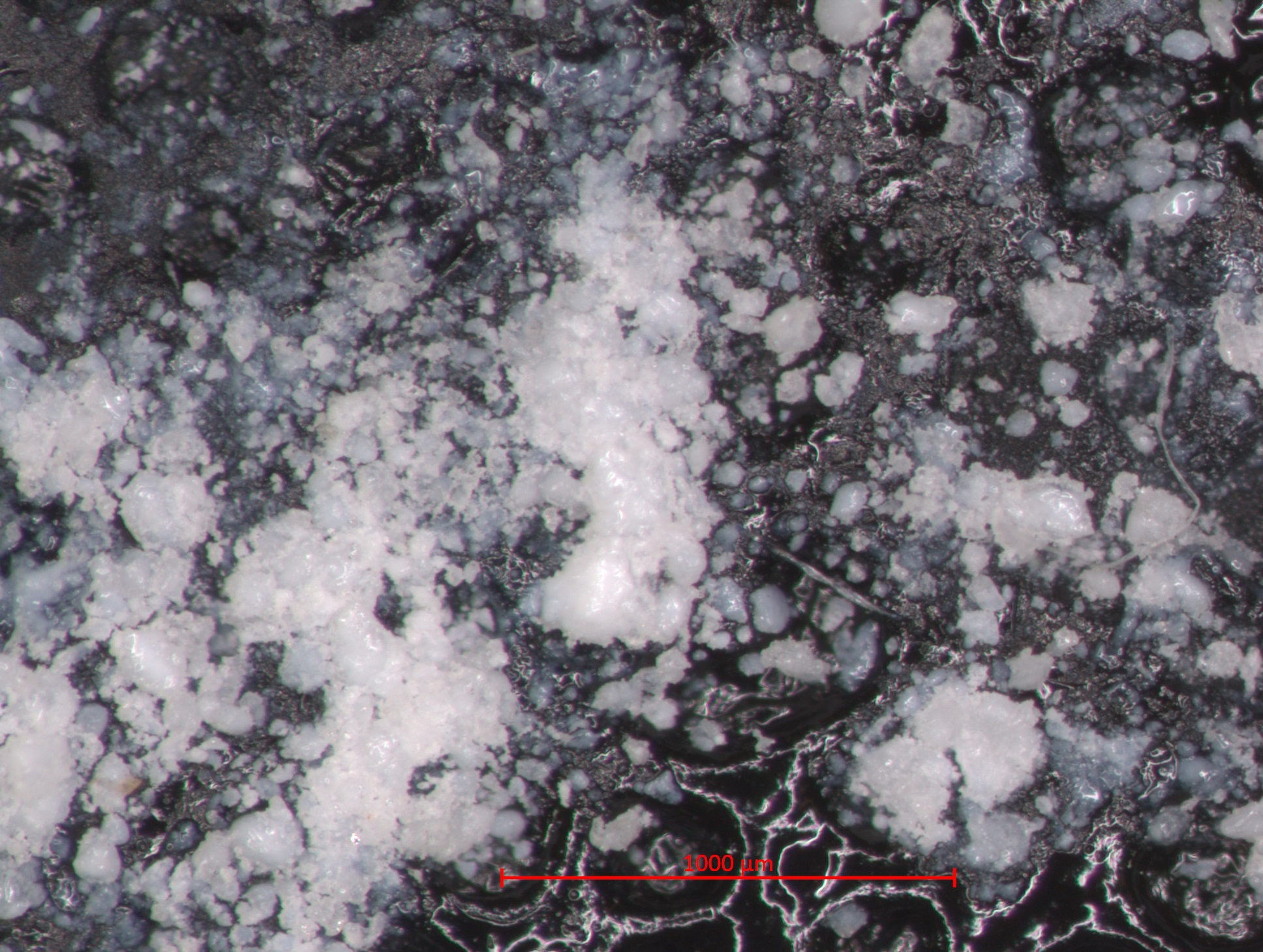
| 13:49, 12 January 2017 | 1 10 2 WhiteSpain STEMI 1000um.jpg (file) |  |
646 KB | Photomicrograph with 1000µm measurement bar (approximate to 50x magnification) (LC) Image captured using Zeiss STEMI SV 11 stereomicroscope. Pigment sample was applied to carbon tape and mounted onto aluminum examination stub. Image was illuminated b... | 1 |
| 13:33, 12 January 2017 | 1 09 11 DryTitaneoc EDS Spectrum.jpg (file) | 82 KB | EDS Spectrum showing elemental peak height as a function of X-ray counts collected (LC) Elements Identified Major (> 10%): barium, oxygen, carbon, titanium. Minor (1-10%): sulfur. Trace (< 1%): NA. Area X-ray counts collected: 1,030,389. Live Time:... | 1 | |
| 13:31, 12 January 2017 | 1 09 11 DryTitaneoc SEM 50um.jpg (file) |  |
600 KB | SEM image with 50µm measurement bar (approximate to 1000x magnification) (LC) Imaged with FEI Quanta 600 scanning electron microscope (SEM) and xT microscope Server user interface. Pigment sample was applied to carbon tape and mounted onto aluminum e... | 1 |
| 13:31, 12 January 2017 | 1 09 11 DryTitaneoc SEM 100um.jpg (file) |  |
665 KB | SEM image with 100µm measurement bar (approximate to 500x magnification) (LC) Imaged with FEI Quanta 600 scanning electron microscope (SEM) and xT microscope Server user interface. Pigment sample was applied to carbon tape and mounted onto aluminum e... | 1 |
| 13:30, 12 January 2017 | 1 09 11 DryTitanoec STEMI 1000um.jpg (file) |  |
764 KB | Photomicrograph with 1000µm measurement bar (approximate to 50x magnification) (LC) Image captured using Zeiss STEMI SV 11 stereomicroscope. Pigment sample was applied to carbon tape and mounted onto aluminum examination stub. Image was illuminated b... | 1 |
| 15:30, 11 January 2017 | 1 09 10 TitaniumWhiteHatfield EDS Spectrum.jpg (file) | 67 KB | EDS Spectrum showing elemental peak height as a function of X-ray counts collected (LC) Elements Identified Major (> 10%): carbon, oxygen, titanium. Minor (1-10%): NA. Trace (< 1%): NA. Area X-ray counts collected: 1,024,473. Live Time: 49.6 second... | 1 | |
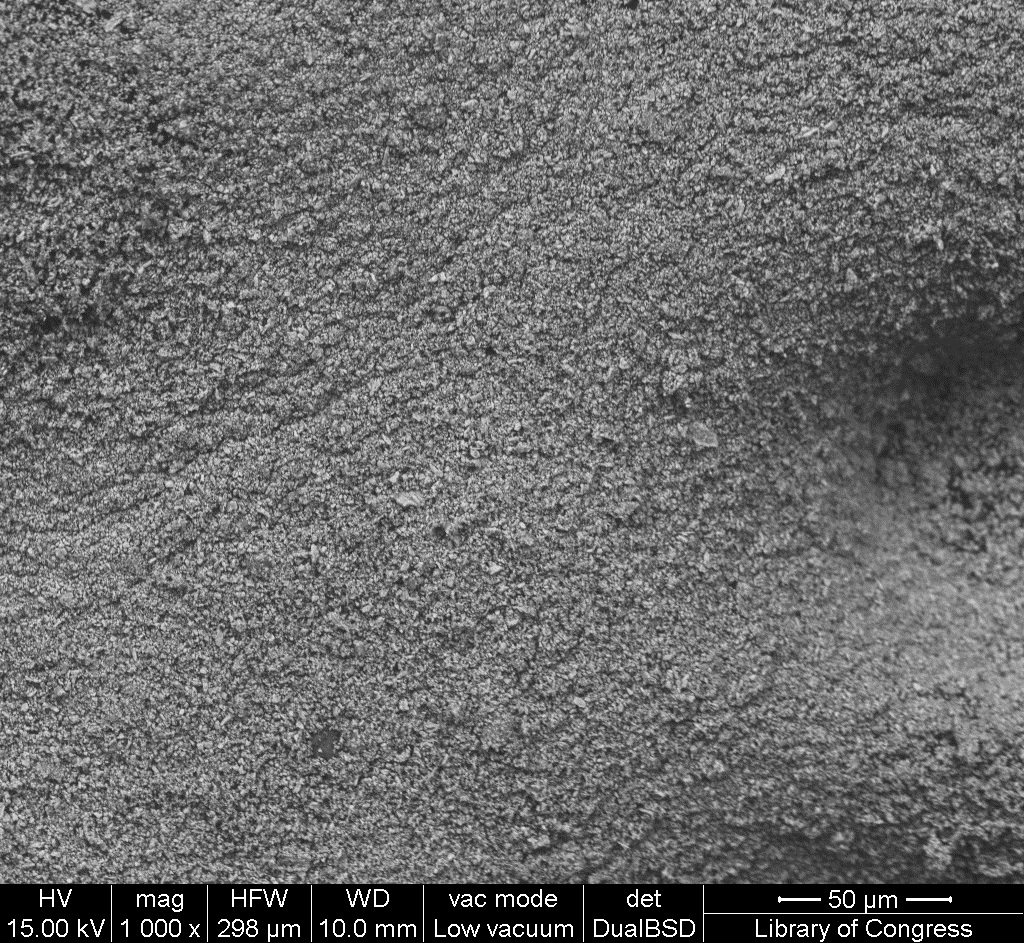
| 15:29, 11 January 2017 | 1 09 10 TitaniumWhiteHatfield SEM 50um.jpg (file) |  |
543 KB | SEM image with 50µm measurement bar (approximate to 1000x magnification) (LC) Imaged with FEI Quanta 600 scanning electron microscope (SEM) and xT microscope Server user interface. Pigment sample was applied to carbon tape and mounted onto aluminum e... | 1 |
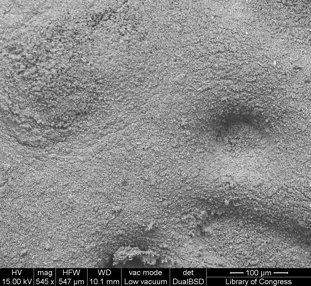
| 15:28, 11 January 2017 | 1 09 10 TitaniumWhiteHatfield SEM 100um.jpg (file) |  |
569 KB | SEM image with 100µm measurement bar (approximate to 500x magnification) (LC) Imaged with FEI Quanta 600 scanning electron microscope (SEM) and xT microscope Server user interface. Pigment sample was applied to carbon tape and mounted onto aluminum e... | 1 |
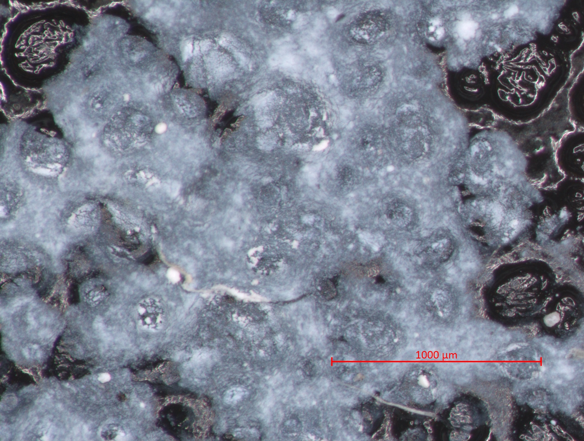
| 15:27, 11 January 2017 | 1 09 10 TitaniumWhiteHatfield STEMI 1000um.jpg (file) |  |
722 KB | Photomicrograph with 1000µm measurement bar (approximate to 50x magnification) (LC) Image captured using Zeiss STEMI SV 11 stereomicroscope. Pigment sample was applied to carbon tape and mounted onto aluminum examination stub. Image was illuminated b... | 1 |
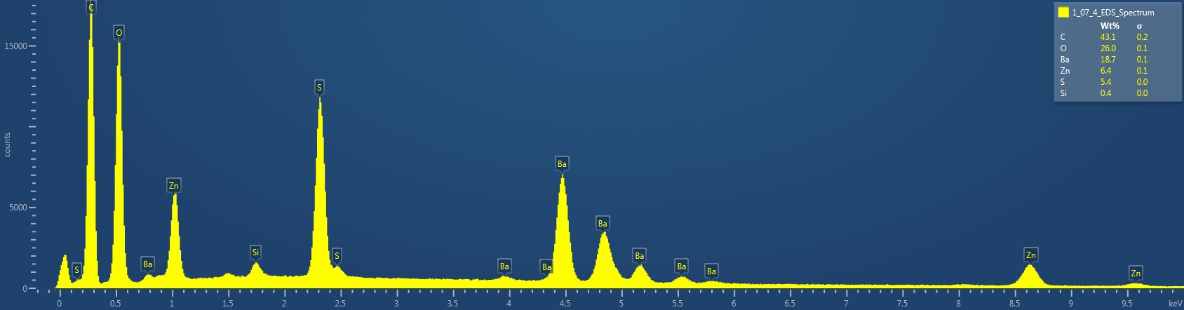
| 15:16, 11 January 2017 | 1 07 4 Lithopone EDS Spectrum.jpg (file) | 85 KB | EDS Spectrum showing elemental peak height as a function of X-ray counts collected (LC) Elements Identified Major (> 10%): carbon, oxygen, barium. Minor (1-10%): zinc, sulfur. Trace (< 1%): silicon. Area X-ray counts collected: 1,021,143. Live Time... | 1 | |
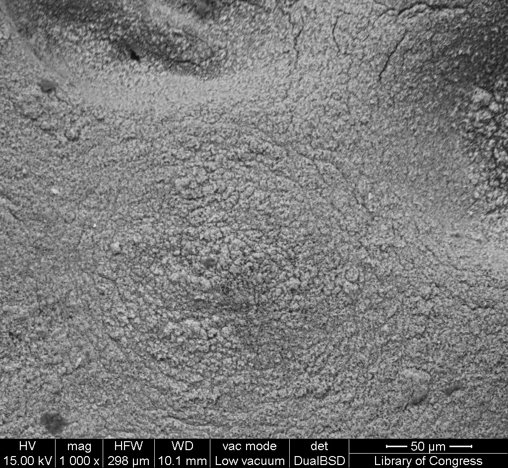
| 15:11, 11 January 2017 | 1 07 4 Lithopone SEM 50um.jpg (file) |  |
543 KB | SEM image with 50µm measurement bar (approximate to 1000x magnification) (LC) Imaged with FEI Quanta 600 scanning electron microscope (SEM) and xT microscope Server user interface. Pigment sample was applied to carbon tape and mounted onto aluminum e... | 1 |
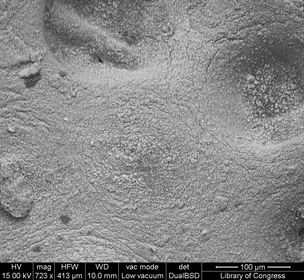
| 15:11, 11 January 2017 | 1 07 4 Lithopone SEM 100um.jpg (file) |  |
610 KB | SEM image with 100µm measurement bar (approximate to 500x magnification) (LC) Imaged with FEI Quanta 600 scanning electron microscope (SEM) and xT microscope Server user interface. Pigment sample was applied to carbon tape and mounted onto aluminum e... | 1 |
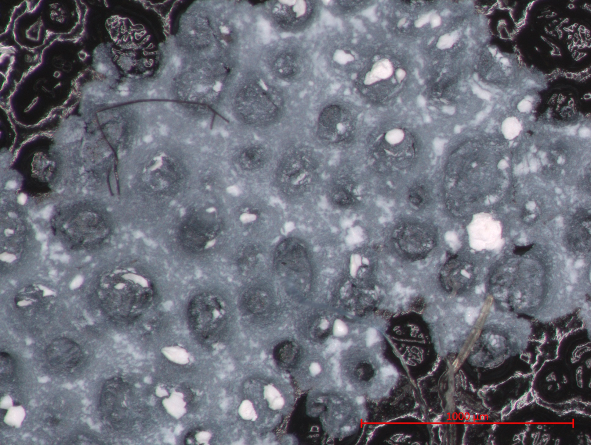
| 15:10, 11 January 2017 | 1 07 4 Lithopone STEMI 1000um.jpg (file) |  |
588 KB | Photomicrograph with 1000µm measurement bar (approximate to 50x magnification) (LC) Image captured using Zeiss STEMI SV 11 stereomicroscope. Pigment sample was applied to carbon tape and mounted onto aluminum examination stub. Image was illuminated b... | 1 |
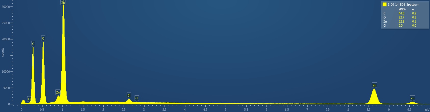
| 14:06, 11 January 2017 | 1 06 14 ZnO EDS Spectrum.jpg (file) | 71 KB | EDS Spectrum showing elemental peak height as a function of X-ray counts collected (LC) Elements Identified Major (> 10%): carbon, oxygen, zinc. Minor (1-10%): NA. Trace (< 1%): chlorine. Area X-ray counts collected: 1,016,534. Live Time: 31.2 seco... | 1 | |
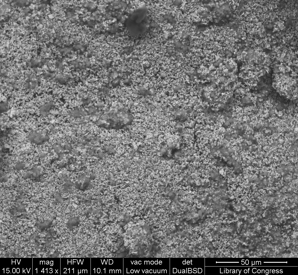
| 14:04, 11 January 2017 | 1 06 14 ZnO SEM 50um.jpg (file) |  |
508 KB | SEM image with 50µm measurement bar (approximate to 1000x magnification) (LC) Imaged with FEI Quanta 600 scanning electron microscope (SEM) and xT microscope Server user interface. Pigment sample was applied to carbon tape and mounted onto aluminum e... | 1 |
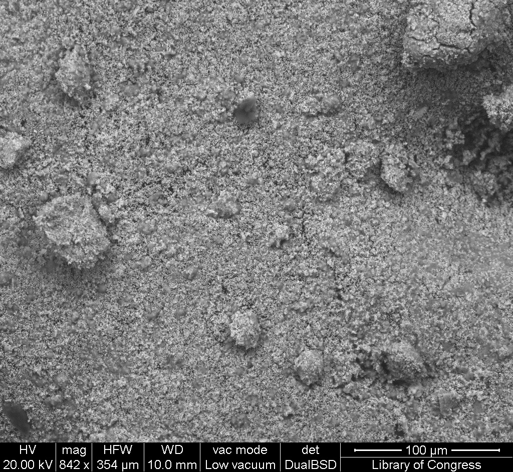
| 14:04, 11 January 2017 | 1 06 14 ZnO SEM 100um.jpg (file) |  |
542 KB | SEM image with 100µm measurement bar (approximate to 500x magnification) (LC) Imaged with FEI Quanta 600 scanning electron microscope (SEM) and xT microscope Server user interface. Pigment sample was applied to carbon tape and mounted onto aluminum e... | 1 |
| 14:02, 11 January 2017 | 1 06 14 ZnO STEMI 1000um.jpg (file) |  |
801 KB | Photomicrograph with 1000µm measurement bar (approximate to 50x magnification) (LC) Image captured using Zeiss STEMI SV 11 stereomicroscope. Pigment sample was applied to carbon tape and mounted onto aluminum examination stub. Image was illuminated b... | 1 |
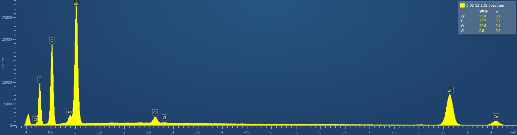
| 13:47, 11 January 2017 | 1 06 13 ZincWhite EDS Spectrum.jpg (file) | 72 KB | EDS Spectrum showing elemental peak height as a function of X-ray counts collected (LC) Elements Identified Major (> 10%): zinc, carbon, oxygen. Minor (1-10%): NA. Trace (< 1%): chlorine. Area X-ray counts collected: 1,024,582. Live Time: 58.3 seco... | 1 | |
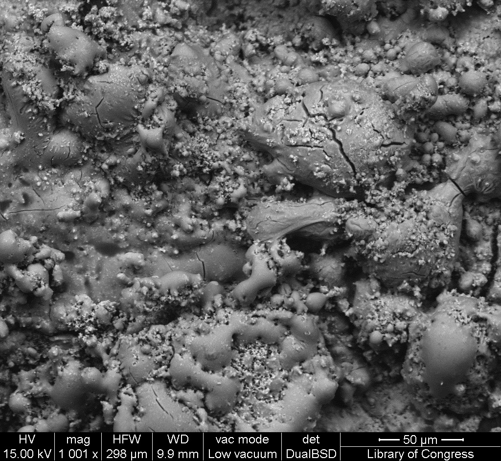
| 13:45, 11 January 2017 | 1 06 13 ZincWhite SEM 50um.jpg (file) |  |
508 KB | SEM image with 50µm measurement bar (approximate to 1000x magnification) (LC) Imaged with FEI Quanta 600 scanning electron microscope (SEM) and xT microscope Server user interface. Pigment sample was applied to carbon tape and mounted onto aluminum e... | 1 |
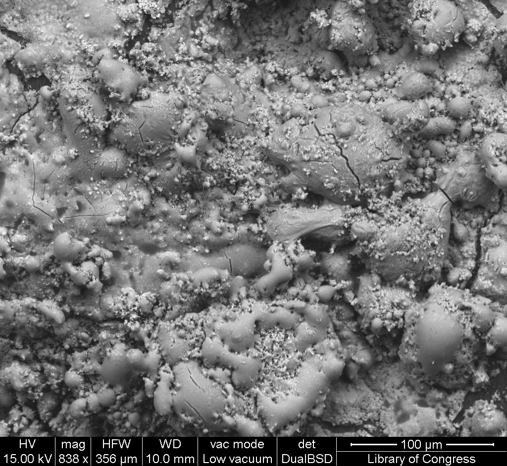
| 13:45, 11 January 2017 | 1 06 13 ZincWhite SEM 100um.jpg (file) |  |
516 KB | SEM image with 100µm measurement bar (approximate to 500x magnification) (LC) Imaged with FEI Quanta 600 scanning electron microscope (SEM) and xT microscope Server user interface. Pigment sample was applied to carbon tape and mounted onto aluminum e... | 1 |
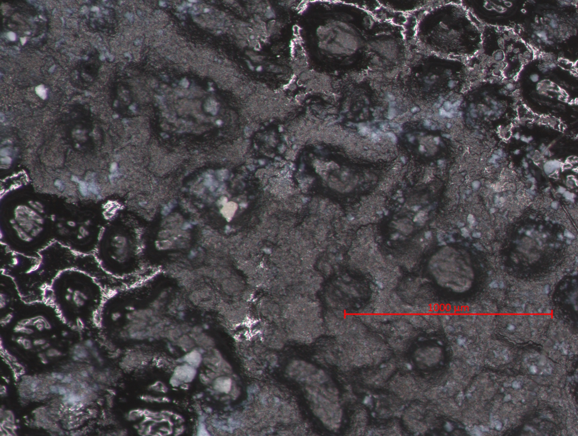
| 13:44, 11 January 2017 | 1 06 13 ZincWhite STEMI 1000um.jpg (file) |  |
802 KB | Photomicrograph with 1000µm measurement bar (approximate to 50x magnification) (LC) Image captured using Zeiss STEMI SV 11 stereomicroscope. Pigment sample was applied to carbon tape and mounted onto aluminum examination stub. Image was illuminated b... | 1 |
| 13:34, 11 January 2017 | 1 06 12 OxideZinc EDS Spectrum.jpg (file) | 74 KB | EDS Spectrum showing elemental peak height as a function of X-ray counts collected (LC) Elements Identified Major (> 10%): zinc, carbon, oxygen. Minor (1-10%): NA. Trace (< 1%): chlorine. Area X-ray counts collected: 1,032,525. Live Time: 57.0 seco... | 1 | |
| 13:32, 11 January 2017 | 1 06 12 OxideZinc SEM 50um.jpg (file) |  |
507 KB | SEM image with 50µm measurement bar (approximate to 1000x magnification) (LC) Imaged with FEI Quanta 600 scanning electron microscope (SEM) and xT microscope Server user interface. Pigment sample was applied to carbon tape and mounted onto aluminum e... | 1 |
| 13:32, 11 January 2017 | 1 06 12 OxideZinc SEM 100um.jpg (file) |  |
547 KB | SEM image with 100µm measurement bar (approximate to 500x magnification) (LC) Imaged with FEI Quanta 600 scanning electron microscope (SEM) and xT microscope Server user interface. Pigment sample was applied to carbon tape and mounted onto aluminum e... | 1 |
| 13:31, 11 January 2017 | 1 06 12 OxideZinc STEMI 1000um.jpg (file) |  |
791 KB | Photomicrograph with 1000µm measurement bar (approximate to 50x magnification) (LC) Image captured using Zeiss STEMI SV 11 stereomicroscope. Pigment sample was applied to carbon tape and mounted onto aluminum examination stub. Image was illuminated b... | 1 |
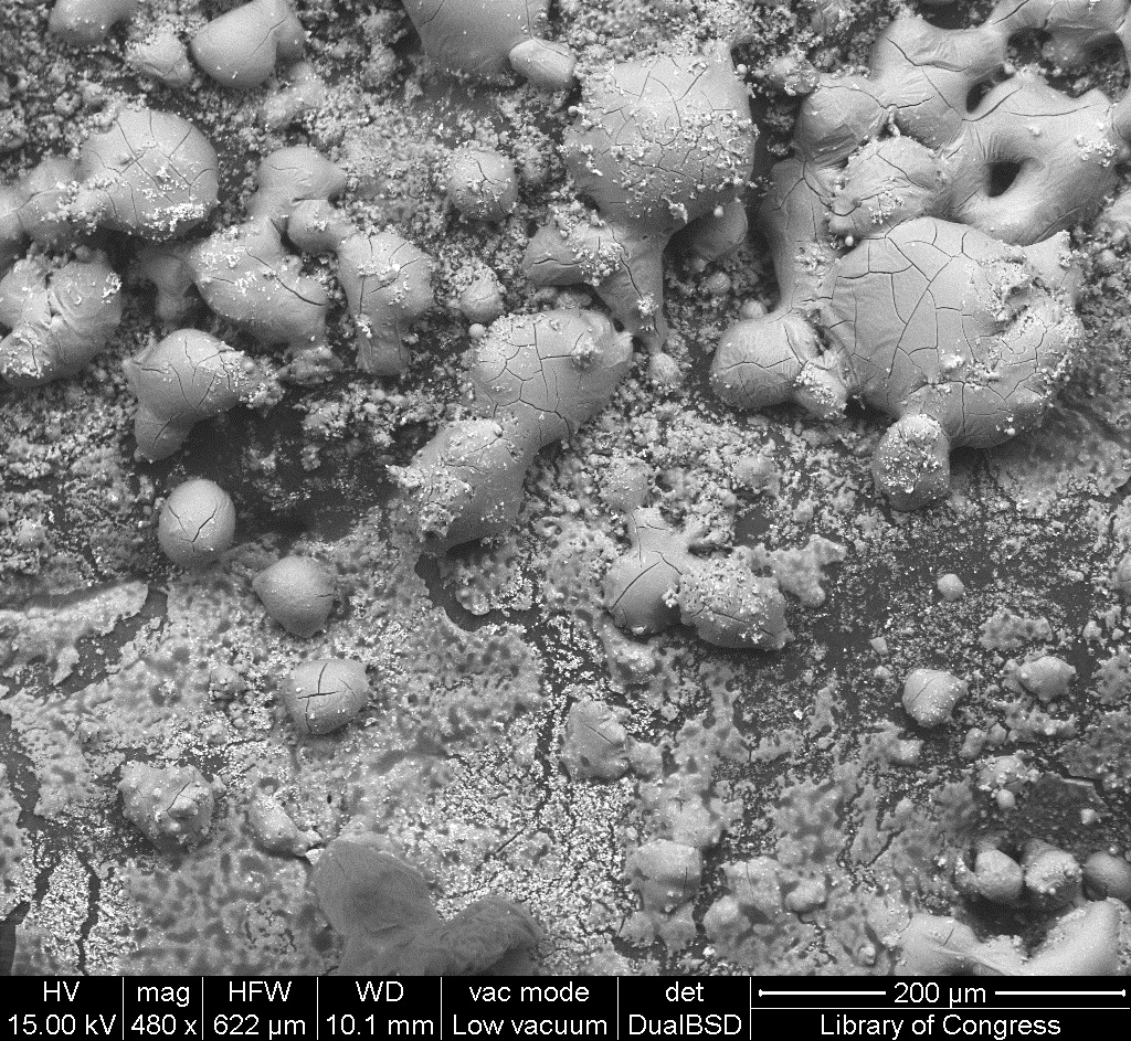
| 12:09, 11 January 2017 | 1 06 11 ZincWhiteWinsorNewton SEM 200um.jpg (file) |  |
575 KB | SEM image with 200µm measurement bar (approximate to 500x magnification) (LC) Imaged with FEI Quanta 600 scanning electron microscope (SEM) and xT microscope Server user interface. Pigment sample was applied to carbon tape and mounted onto aluminum e... | 1 |
| 12:01, 11 January 2017 | 1 06 11 ZincWhiteWinsorNewton EDS Spectrum.jpg (file) | 79 KB | EDS Spectrum showing elemental peak height as a function of X-ray counts collected (LC) Elements Identified Major (> 10%): carbon, oxygen, zinc. Minor (1-10%): NA. Trace (< 1%): chlorine, barium, titanium, sulfur. Area X-ray counts collected: 1,022... | 1 | |
| 11:59, 11 January 2017 | 1 06 11 ZincWhiteWinsorNewton SEM 50um.jpg (file) |  |
486 KB | SEM image with 500µm measurement bar (approximate to 100x magnification) (LC) Imaged with FEI Quanta 600 scanning electron microscope (SEM) and xT microscope Server user interface. Pigment sample was applied to carbon tape and mounted onto aluminum e... | 1 |
| 11:58, 11 January 2017 | 1 06 11 ZincWhiteWinsorNewton SEM 100um.jpg (file) |  |
575 KB | SEM image with 100µm measurement bar (approximate to 500x magnification) (LC) Imaged with FEI Quanta 600 scanning electron microscope (SEM) and xT microscope Server user interface. Pigment sample was applied to carbon tape and mounted onto aluminum e... | 1 |
| 11:57, 11 January 2017 | 1 06 11 ZincWhiteWinsorNewton STEMI 1000um.jpg (file) |  |
858 KB | Photomicrograph with 1000µm measurement bar (approximate to 50x magnification) (LC) Image captured using Zeiss STEMI SV 11 stereomicroscope. Pigment sample was applied to carbon tape and mounted onto aluminum examination stub. Image was illuminated b... | 1 |
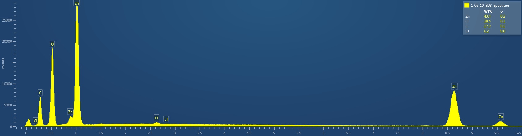
| 11:15, 11 January 2017 | 1 06 10 ZincOxideBaker EDS Spectrum.jpg (file) | 72 KB | EDS Spectrum showing elemental peak height as a function of X-ray counts collected (LC) Elements Identified Major (> 10%): zinc, oxygen, carbon. Minor (1-10%): NA. Trace (< 1%): chlorine. Area X-ray counts collected: 1,018,100. Live Time: 36.2 seco... | 1 | |
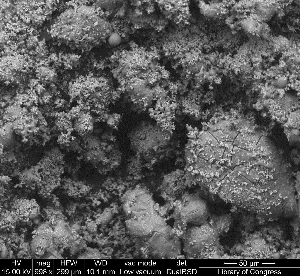
| 11:14, 11 January 2017 | 1 06 10 ZincOxideBaker SEM 50um.jpg (file) |  |
512 KB | SEM image with 500µm measurement bar (approximate to 1000x magnification) (LC) Imaged with FEI Quanta 600 scanning electron microscope (SEM) and xT microscope Server user interface. Pigment sample was applied to carbon tape and mounted onto aluminum ... | 1 |
| 11:13, 11 January 2017 | 1 06 10 ZincOxideBaker SEM 100um.jpg (file) |  |
573 KB | SEM image with 100µm measurement bar (approximate to 500x magnification) (LC) Imaged with FEI Quanta 600 scanning electron microscope (SEM) and xT microscope Server user interface. Pigment sample was applied to carbon tape and mounted onto aluminum e... | 1 |
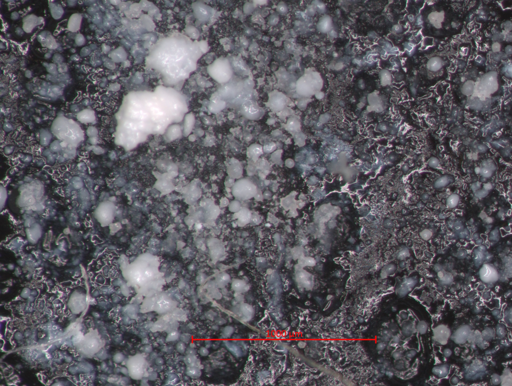
| 11:12, 11 January 2017 | 1 06 10 ZincOxideBaker STEMI 1000um.jpg (file) |  |
711 KB | Photomicrograph with 1000µm measurement bar (approximate to 50x magnification) (LC) Image captured using Zeiss STEMI SV 11 stereomicroscope. Pigment sample was applied to carbon tape and mounted onto aluminum examination stub. Image was illuminated b... | 1 |
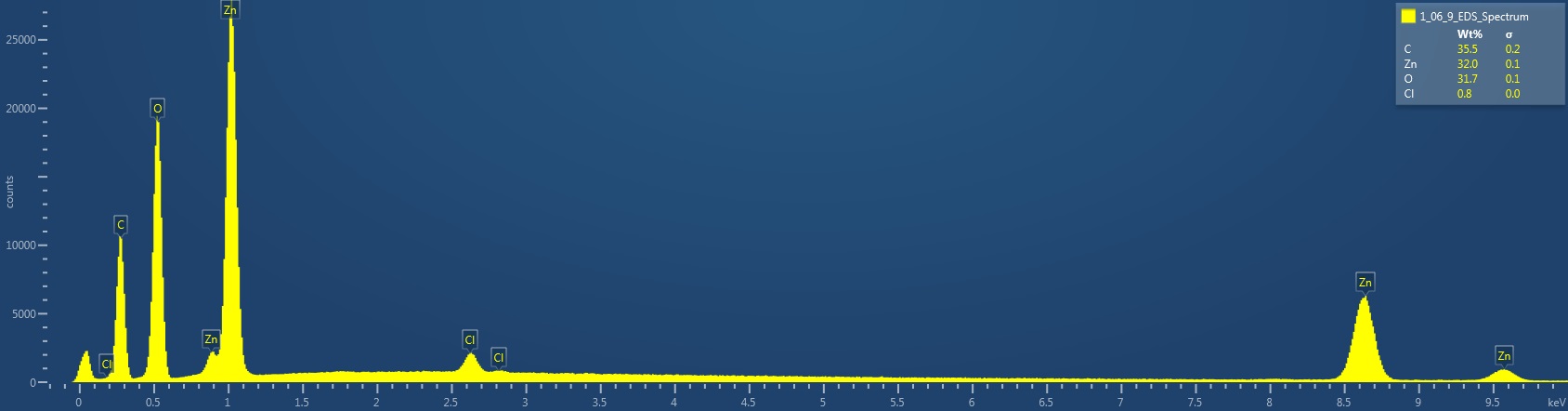
| 10:53, 11 January 2017 | 1 06 9 ZincOxide EDS Spectrum.jpg (file) | 72 KB | EDS Spectrum showing elemental peak height as a function of X-ray counts collected (LC) Elements Identified Major (> 10%): carbon, zinc, oxygen. Minor (1-10%): NA. Trace (< 1%): chlorine. Area X-ray counts collected: 1,021,534. Live Time: 50.3 seco... | 1 | |
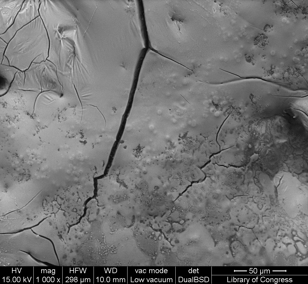
| 10:52, 11 January 2017 | 1 06 9 ZincOxide SEM 50um.jpg (file) |  |
445 KB | SEM image with 50µm measurement bar (approximate to 1000x magnification) (LC) Imaged with FEI Quanta 600 scanning electron microscope (SEM) and xT microscope Server user interface. Pigment sample was applied to carbon tape and mounted onto aluminum e... | 1 |
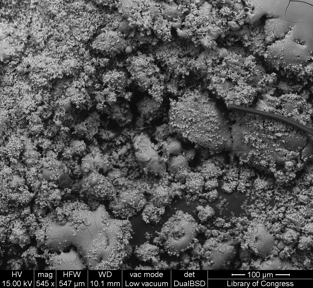
| 10:51, 11 January 2017 | 1 06 9 ZincOxide SEM 100um.jpg (file) |  |
461 KB | SEM image with 100µm measurement bar (approximate to 500x magnification) (LC) Imaged with FEI Quanta 600 scanning electron microscope (SEM) and xT microscope Server user interface. Pigment sample was applied to carbon tape and mounted onto aluminum e... | 1 |
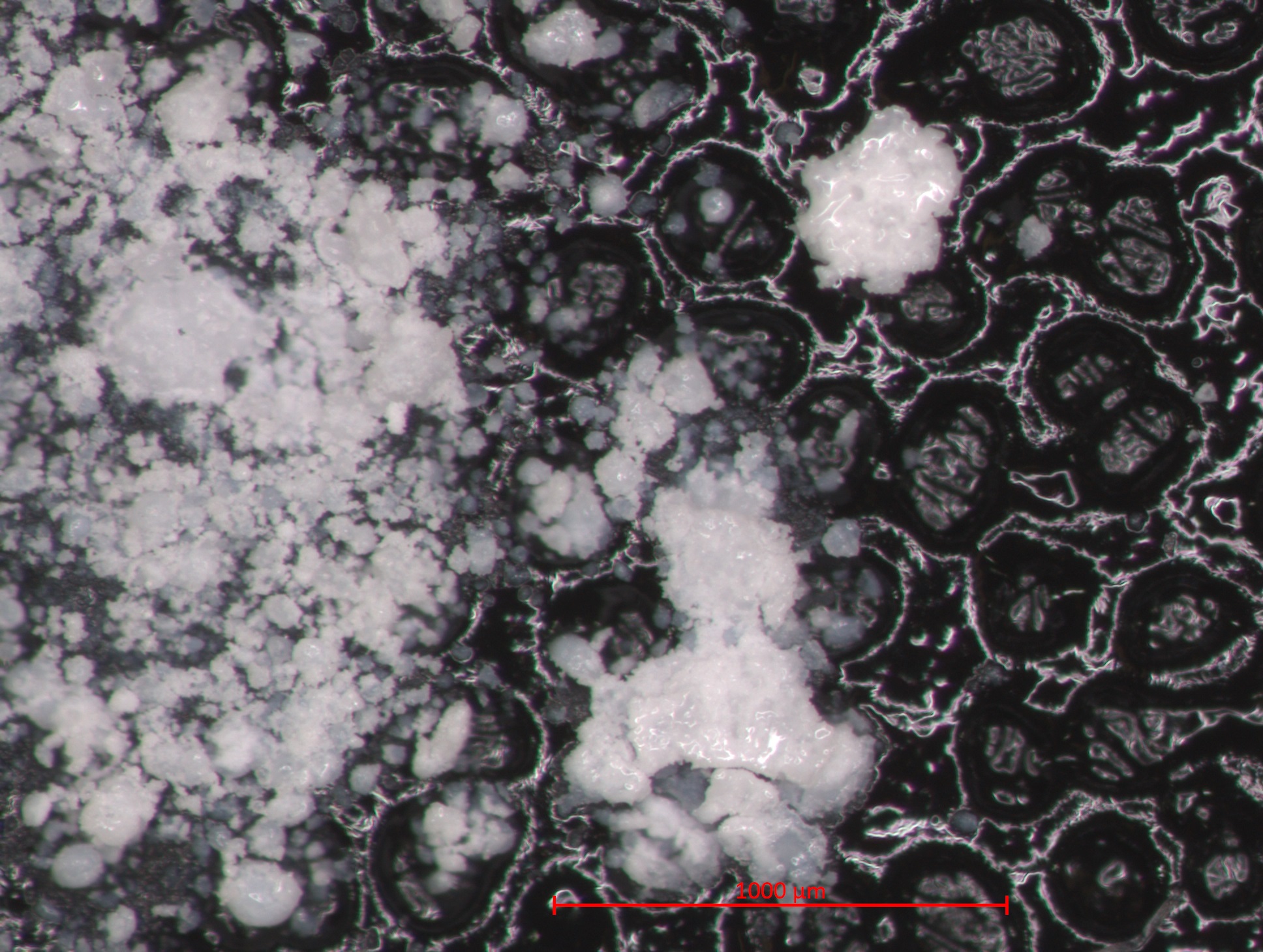
| 10:50, 11 January 2017 | 1 06 9 ZincOxide STEMI 1000um.jpg (file) |  |
869 KB | Photomicrograph with 1000µm measurement bar (approximate to 50x magnification) (LC) Image captured using Zeiss STEMI SV 11 stereomicroscope. Pigment sample was applied to carbon tape and mounted onto aluminum examination stub. Image was illuminated b... | 1 |
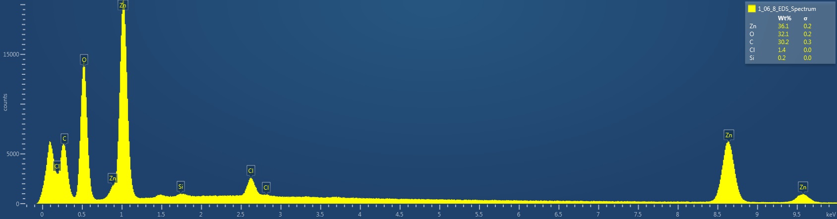
| 10:09, 11 January 2017 | 1 06 8 PermanentChineseWhite EDS Spectrum.jpg (file) | 77 KB | EDS Spectrum showing elemental peak height as a function of X-ray counts collected (LC) Elements Identified Major (> 10%): zinc, oxygen, carbon. Minor (1-10%): chlorine. Trace (< 1%): silicon. Area X-ray counts collected: 1,025,032. Live Time: 42.7... | 1 | |
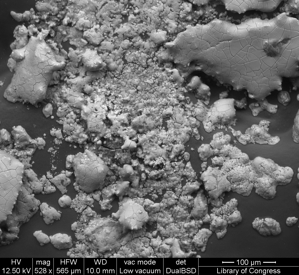
| 10:08, 11 January 2017 | 1 06 8 PermanentChineseWhite SEM 50um.jpg (file) |  |
456 KB | SEM image with 50µm measurement bar (approximate to 1000x magnification) (LC) Imaged with FEI Quanta 600 scanning electron microscope (SEM) and xT microscope Server user interface. Pigment sample was applied to carbon tape and mounted onto aluminum e... | 1 |
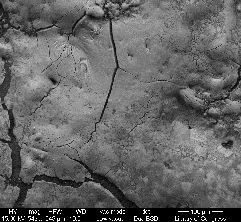
| 10:07, 11 January 2017 | 1 06 8 PermanentChineseWhite SEM 100um.jpg (file) |  |
483 KB | SEM image with 100µm measurement bar (approximate to 500x magnification) (LC) Imaged with FEI Quanta 600 scanning electron microscope (SEM) and xT microscope Server user interface. Pigment sample was applied to carbon tape and mounted onto aluminum e... | 1 |
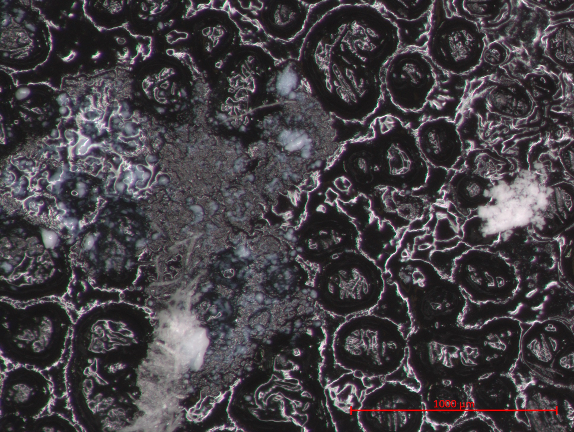
| 10:06, 11 January 2017 | 1 06 8 PermanentChineseWhite STEMI 1000um.jpg (file) |  |
597 KB | Photomicrograph with 1000µm measurement bar (approximate to 50x magnification) (LC) Image captured using Zeiss STEMI SV 11 stereomicroscope. Pigment sample was applied to carbon tape and mounted onto aluminum examination stub. Image was illuminated b... | 1 |
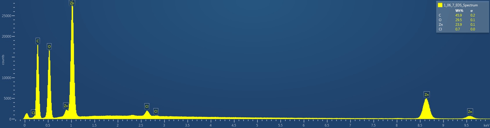
| 09:46, 11 January 2017 | 1 06 7 GreenSeal EDS Spectrum.jpg (file) | 72 KB | EDS Spectrum showing elemental peak height as a function of X-ray counts collected (LC) Elements Identified Major (> 10%): carbon, oxygen, zinc. Minor (1-10%): NA. Trace (< 1%): chlorine. Area X-ray counts collected: 1,016,148. Live Time: 32.6 seco... | 1 | |
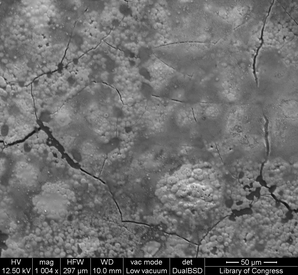
| 09:44, 11 January 2017 | 1 06 7 GreenSeal SEM 50um.jpg (file) |  |
430 KB | SEM image with 50µm measurement bar (approximate to 1000x magnification) (LC) Imaged with FEI Quanta 600 scanning electron microscope (SEM) and xT microscope Server user interface. Pigment sample was applied to carbon tape and mounted onto aluminum e... | 1 |
| 09:43, 11 January 2017 | 1 06 7 GreenSeal SEM 100um.jpg (file) |  |
455 KB | SEM image with 100µm measurement bar (approximate to 500x magnification) (LC) Imaged with FEI Quanta 600 scanning electron microscope (SEM) and xT microscope Server user interface. Pigment sample was applied to carbon tape and mounted onto aluminum e... | 1 |
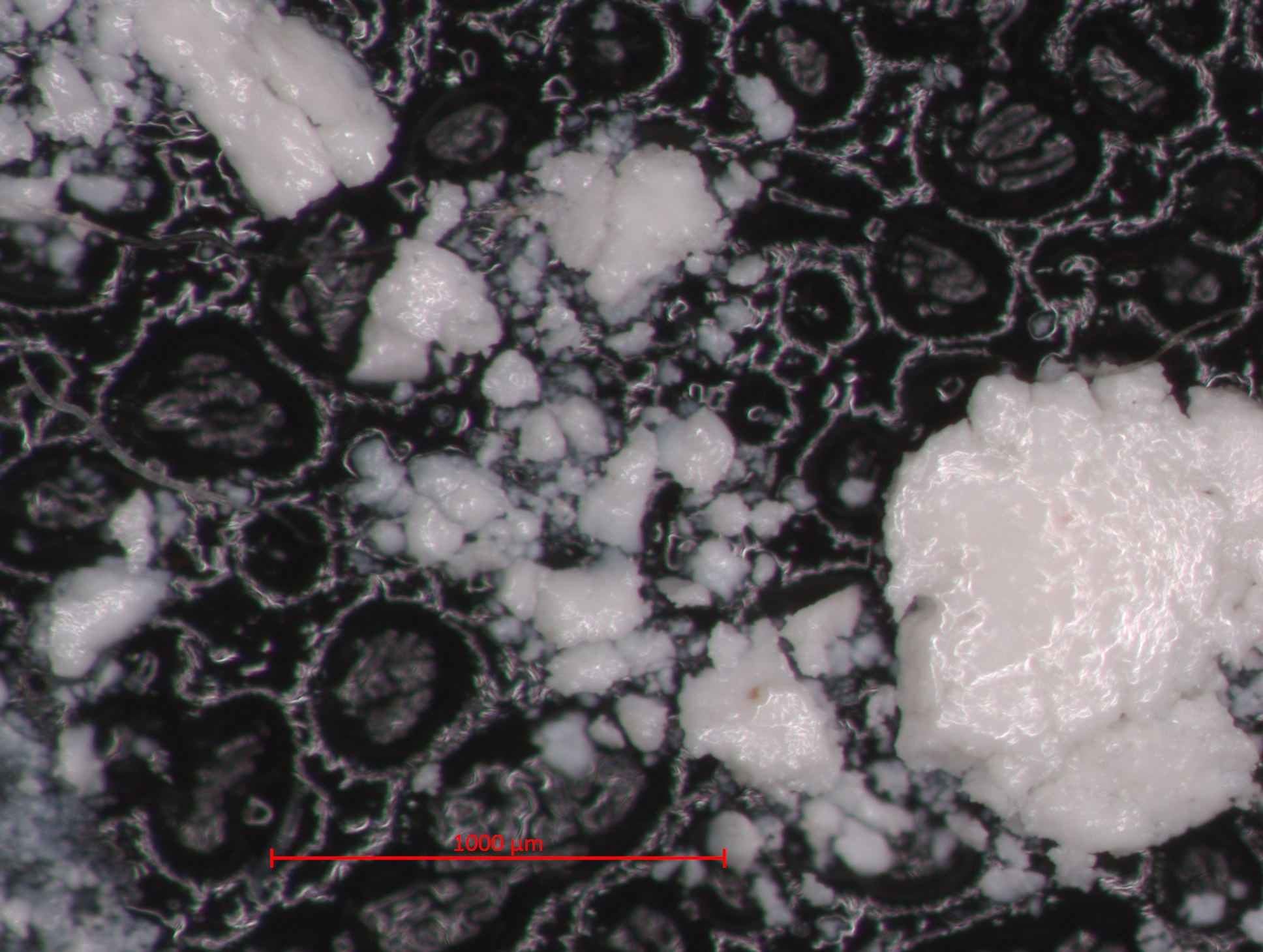
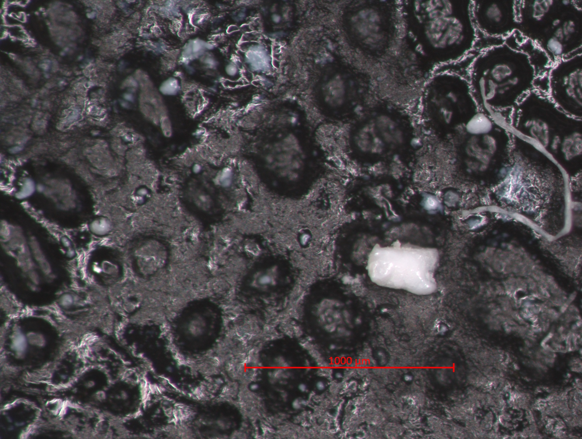
| 09:42, 11 January 2017 | 1 06 7 GreenSeal STEMI 1000um.jpg (file) |  |
725 KB | Photomicrograph with 1000µm measurement bar (approximate to 50x magnification) (LC) Image captured using Zeiss STEMI SV 11 stereomicroscope. Pigment sample was applied to carbon tape and mounted onto aluminum examination stub. Image was illuminated b... | 1 |
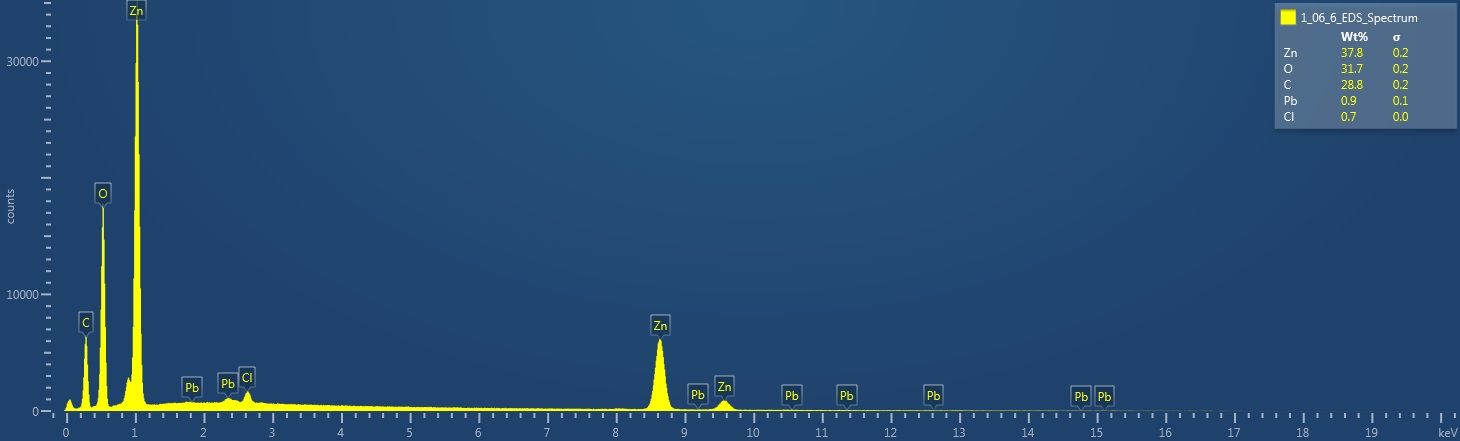
| 15:07, 10 January 2017 | 1 06 6 ZincWhite EDS Spectrum.jpg (file) |  |
61 KB | EDS Spectrum showing elemental peak height as a function of X-ray counts collected (LC) Elements Identified Major (> 10%): zinc, oxygen, carbon. Minor (1-10%): NA. Trace (< 1%): lead, chlorine. Area X-ray counts collected: 1,011,879. Live Time: 24.... | 1 |
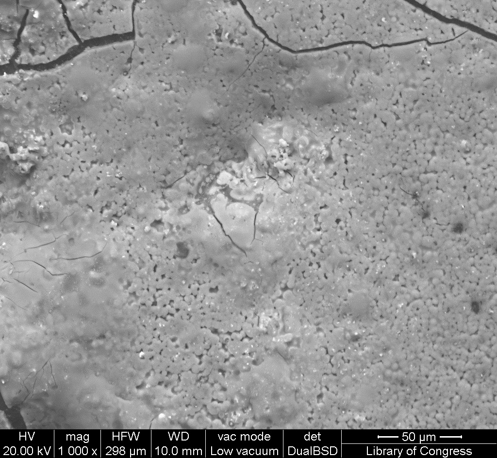
| 15:05, 10 January 2017 | 1 06 6 ZincWhite SEM 50um.jpg (file) |  |
445 KB | SEM image with 50µm measurement bar (approximate to 1000x magnification) (LC) Imaged with FEI Quanta 600 scanning electron microscope (SEM) and xT microscope Server user interface. Pigment sample was applied to carbon tape and mounted onto aluminum e... | 1 |