Uploads by MKullman
Jump to navigation
Jump to search
This special page shows all uploaded files.
| Date | Name | Thumbnail | Size | Description | Versions |
|---|---|---|---|---|---|
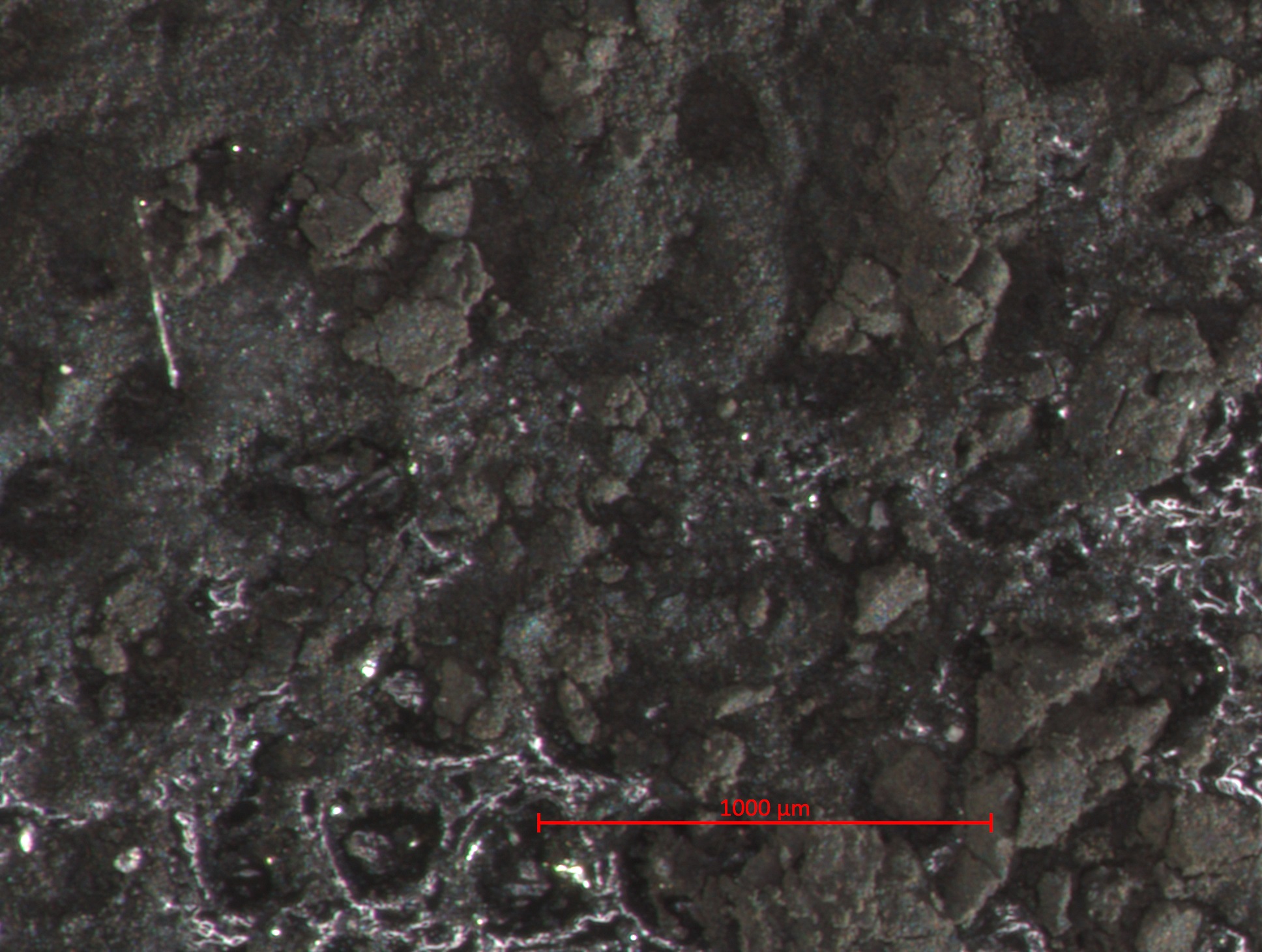
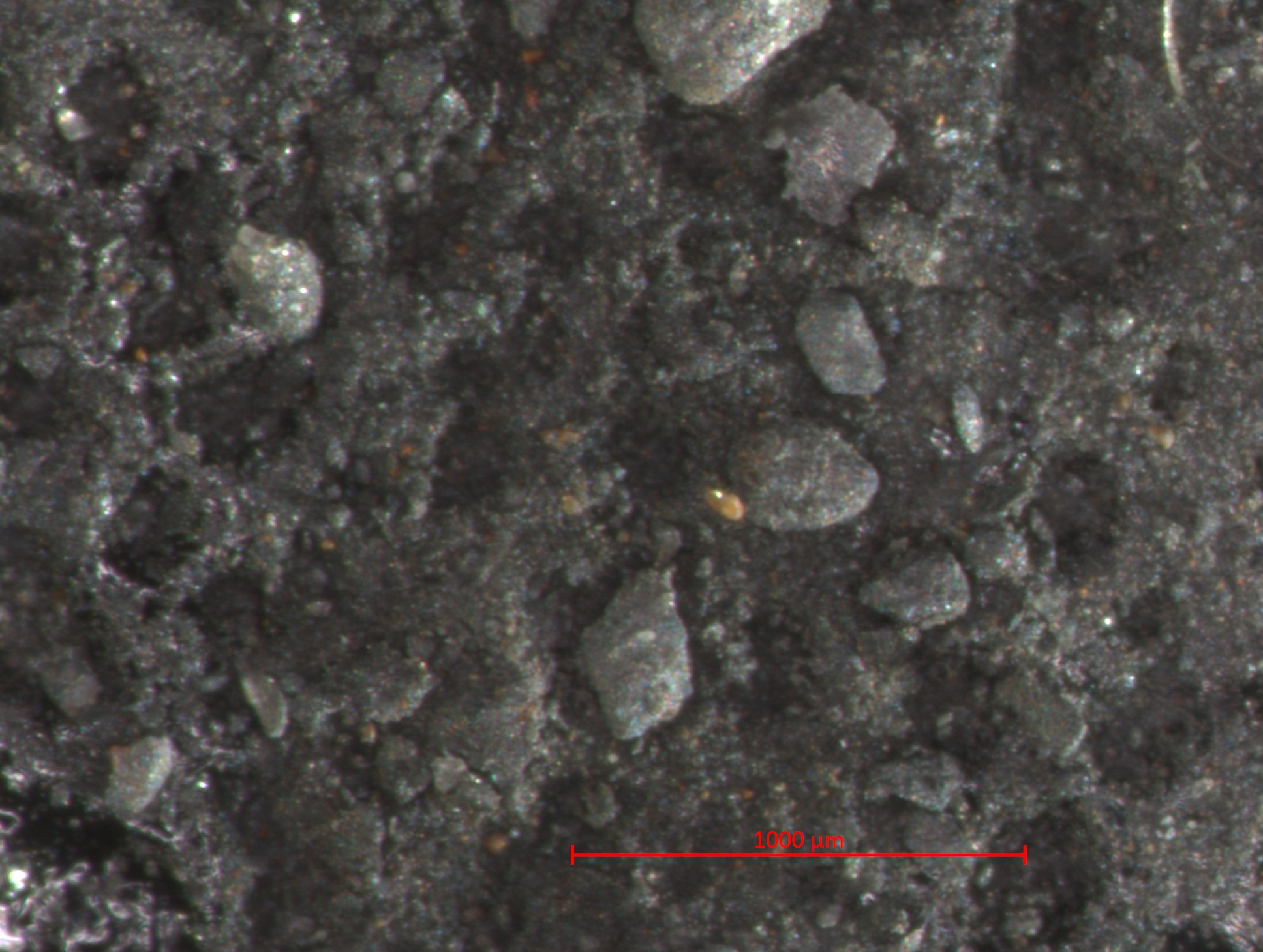
| 15:18, 26 January 2017 | 2 02 7 IvoryBlackRoberson STEMI 1000um.jpg (file) |  |
602 KB | Photomicrograph with 1000µm measurement bar (approximate to 50x magnification) (LC) Image captured using Zeiss STEMI SV 11 stereomicroscope. Pigment sample was applied to carbon tape and mounted onto aluminum examination stub. Image was illuminated b... | 1 |
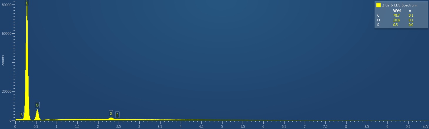
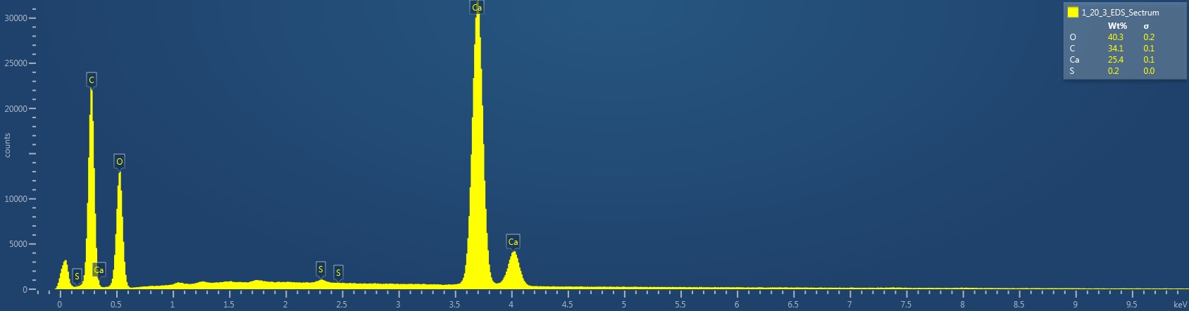
| 15:07, 26 January 2017 | 2 02 6 IvoryBlackWeber EDS Spectrum.jpg (file) |  |
53 KB | EDS Spectrum showing elemental peak height as a function of X-ray counts collected (LC) Elements Identified Major (> 10%): carbon, oxygen. Minor (1-10%): NA. Trace (< 1%): sulfur. Area X-ray counts collected: 1,017,081. Live Time: 21.4 seconds. Mag... | 1 |
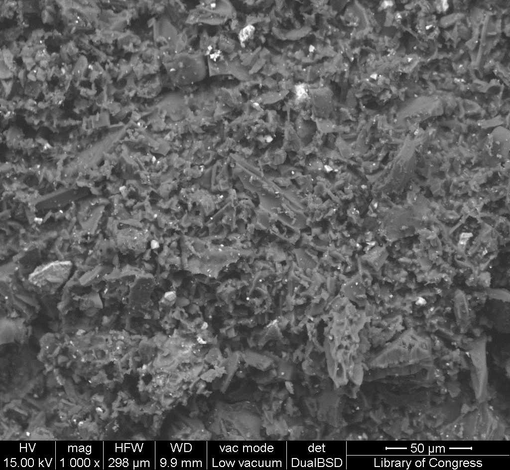
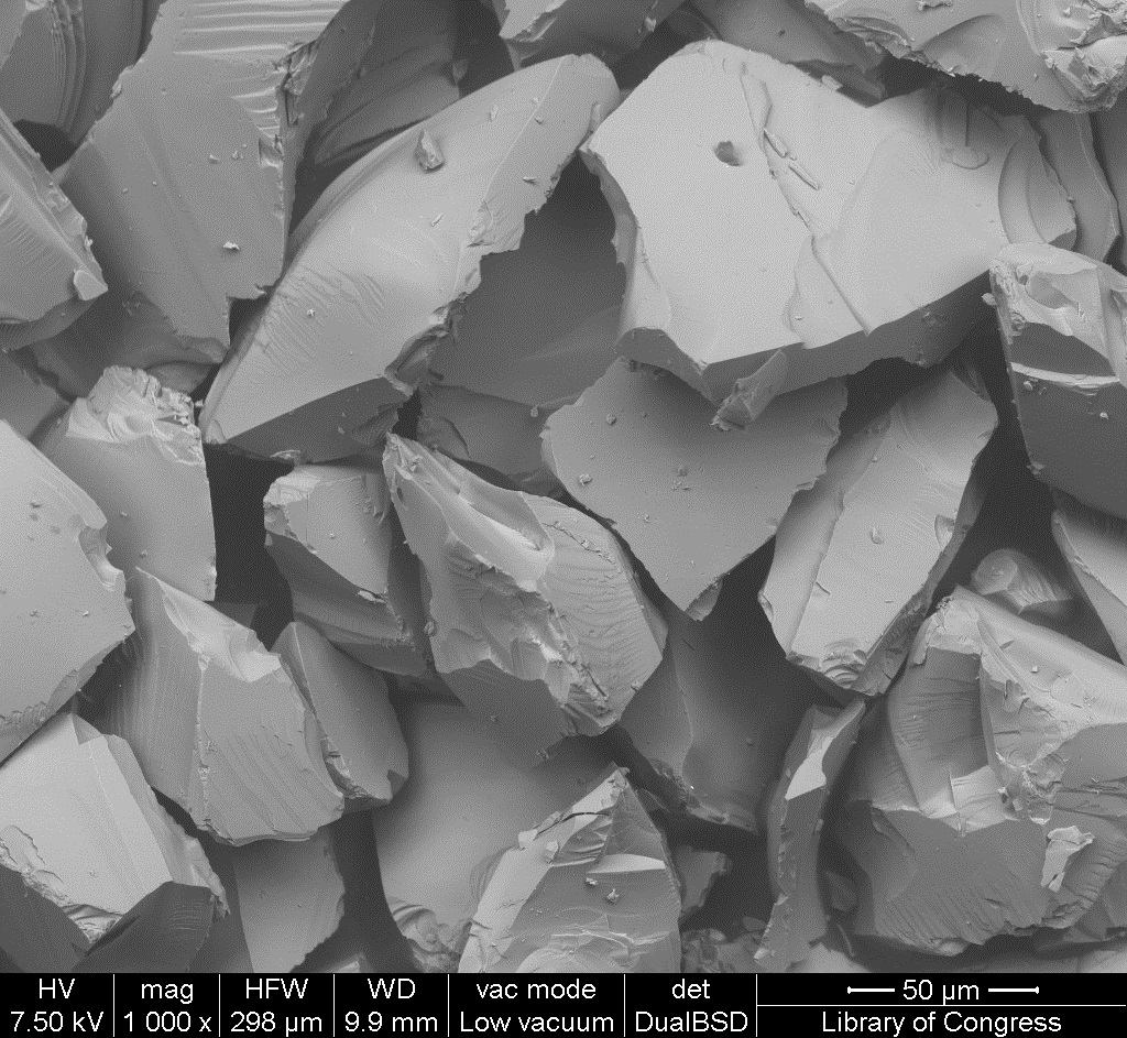
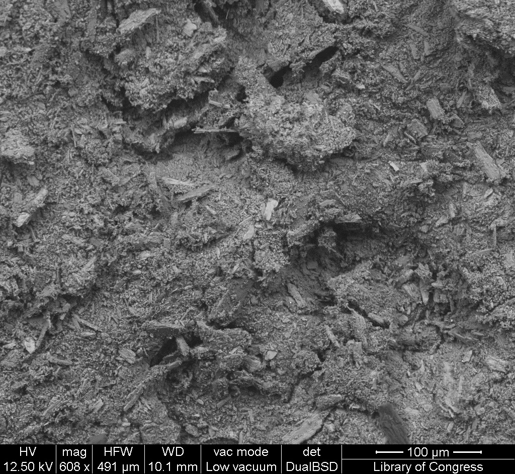
| 15:05, 26 January 2017 | 2 02 6 IvoryBlackWeber SEM 50um.jpg (file) |  |
497 KB | SEM image with 50µm measurement bar (approximate to 1000x magnification) (LC) Imaged with FEI Quanta 600 scanning electron microscope (SEM) and xT microscope Server user interface. Pigment sample was applied to carbon tape and mounted onto aluminum e... | 1 |
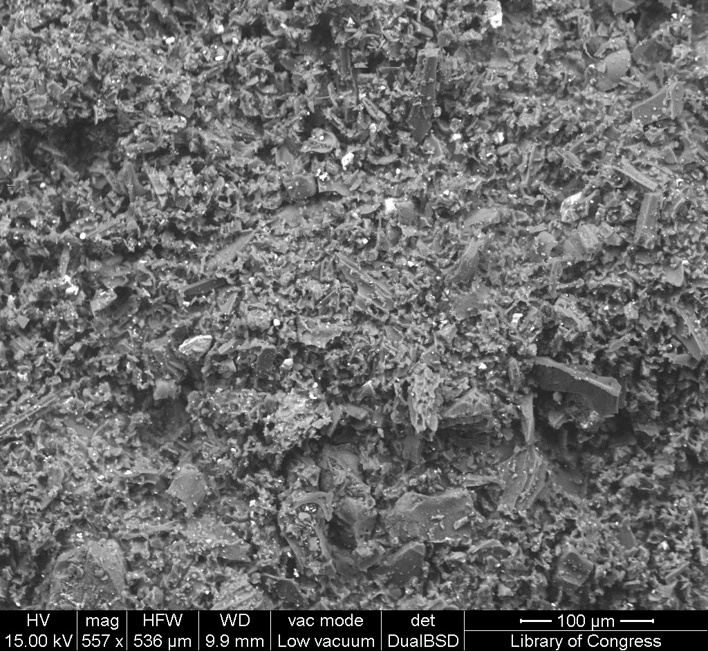
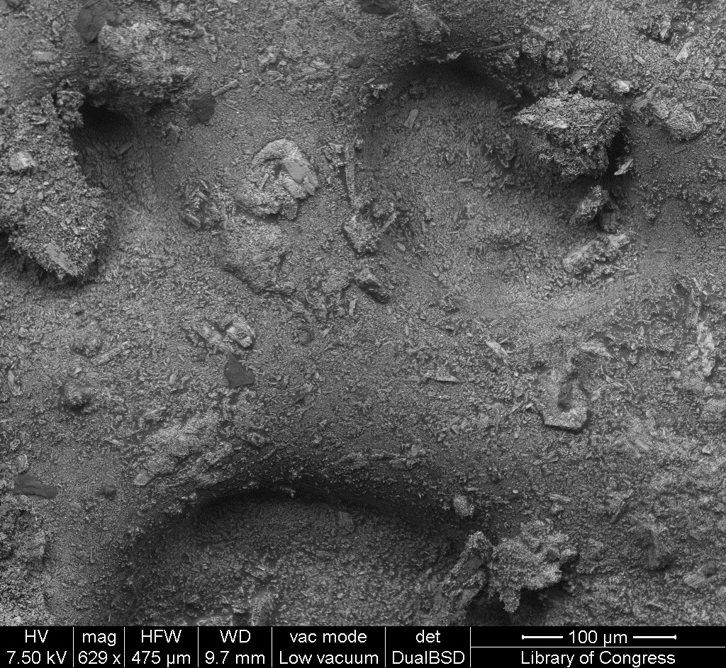
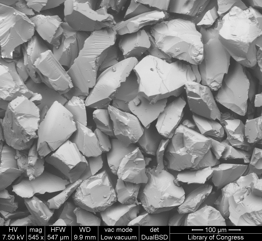
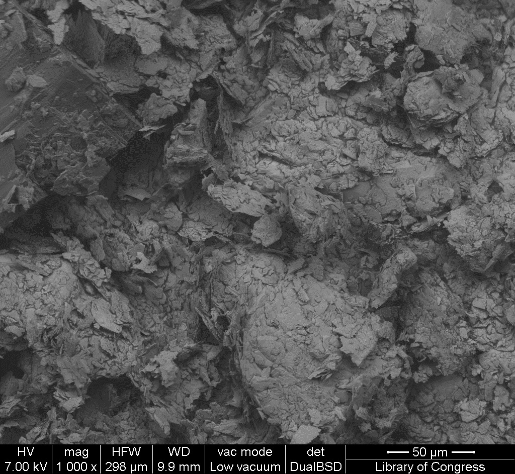
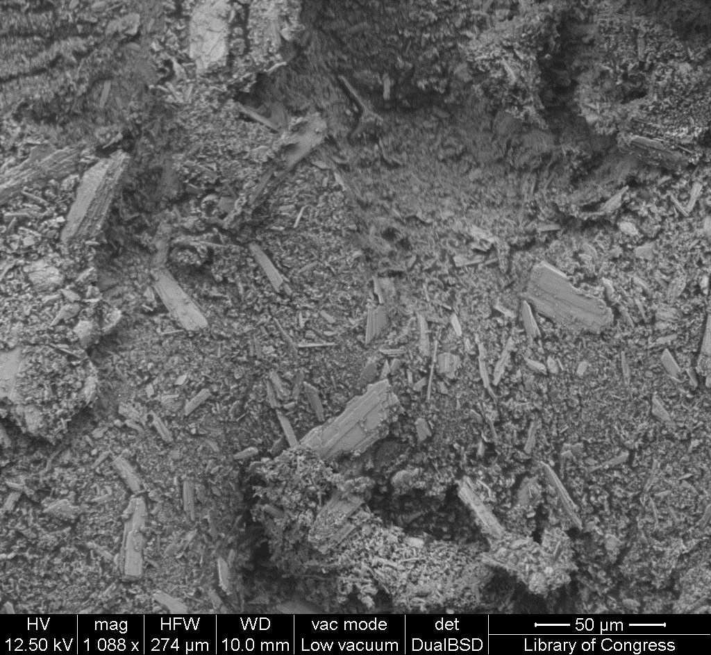
| 15:04, 26 January 2017 | 2 02 6 IvoryBlackWeber SEM 100um.jpg (file) |  |
529 KB | SEM image with 100µm measurement bar (approximate to 500x magnification) (LC) Imaged with FEI Quanta 600 scanning electron microscope (SEM) and xT microscope Server user interface. Pigment sample was applied to carbon tape and mounted onto aluminum e... | 1 |
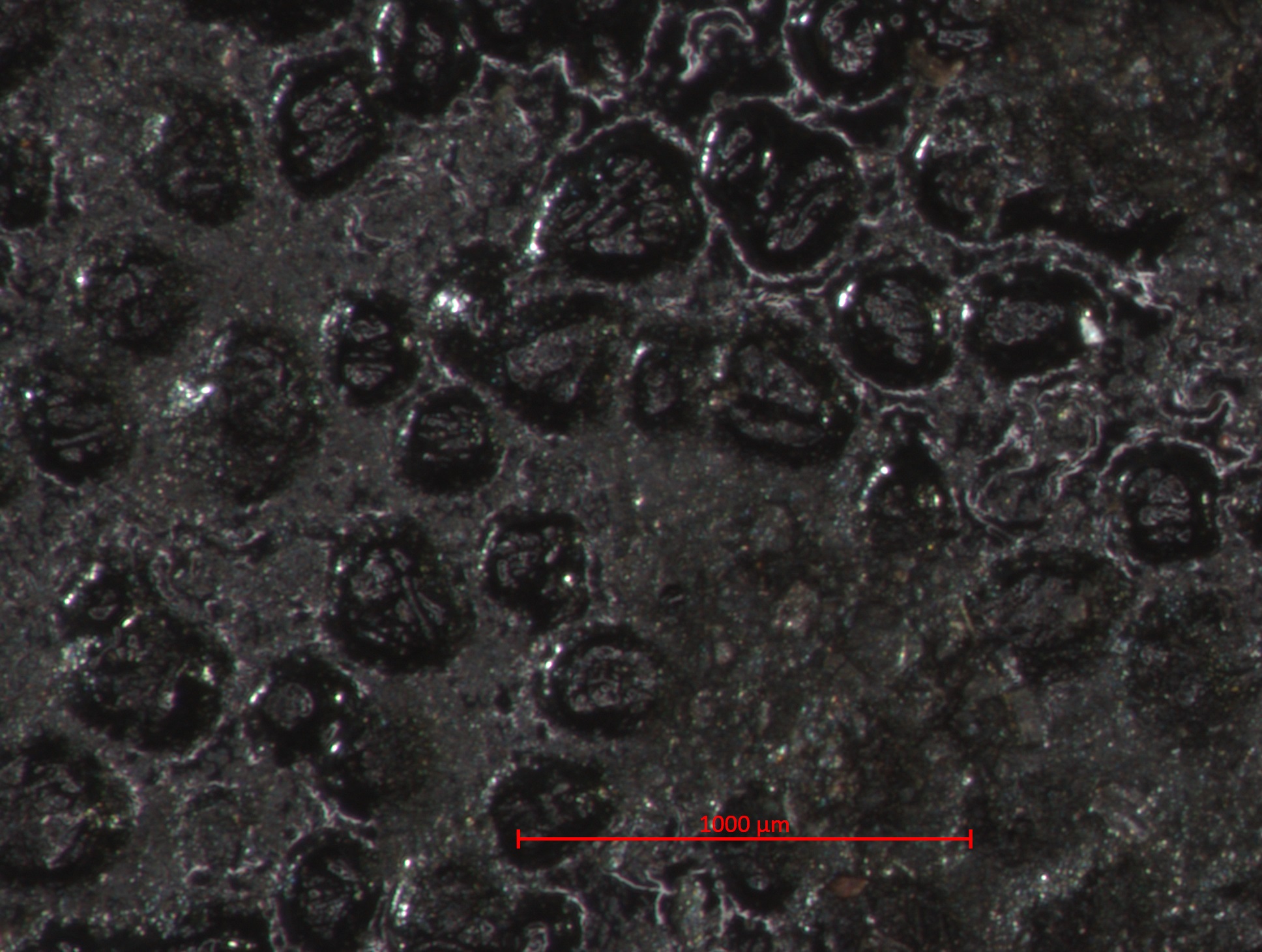
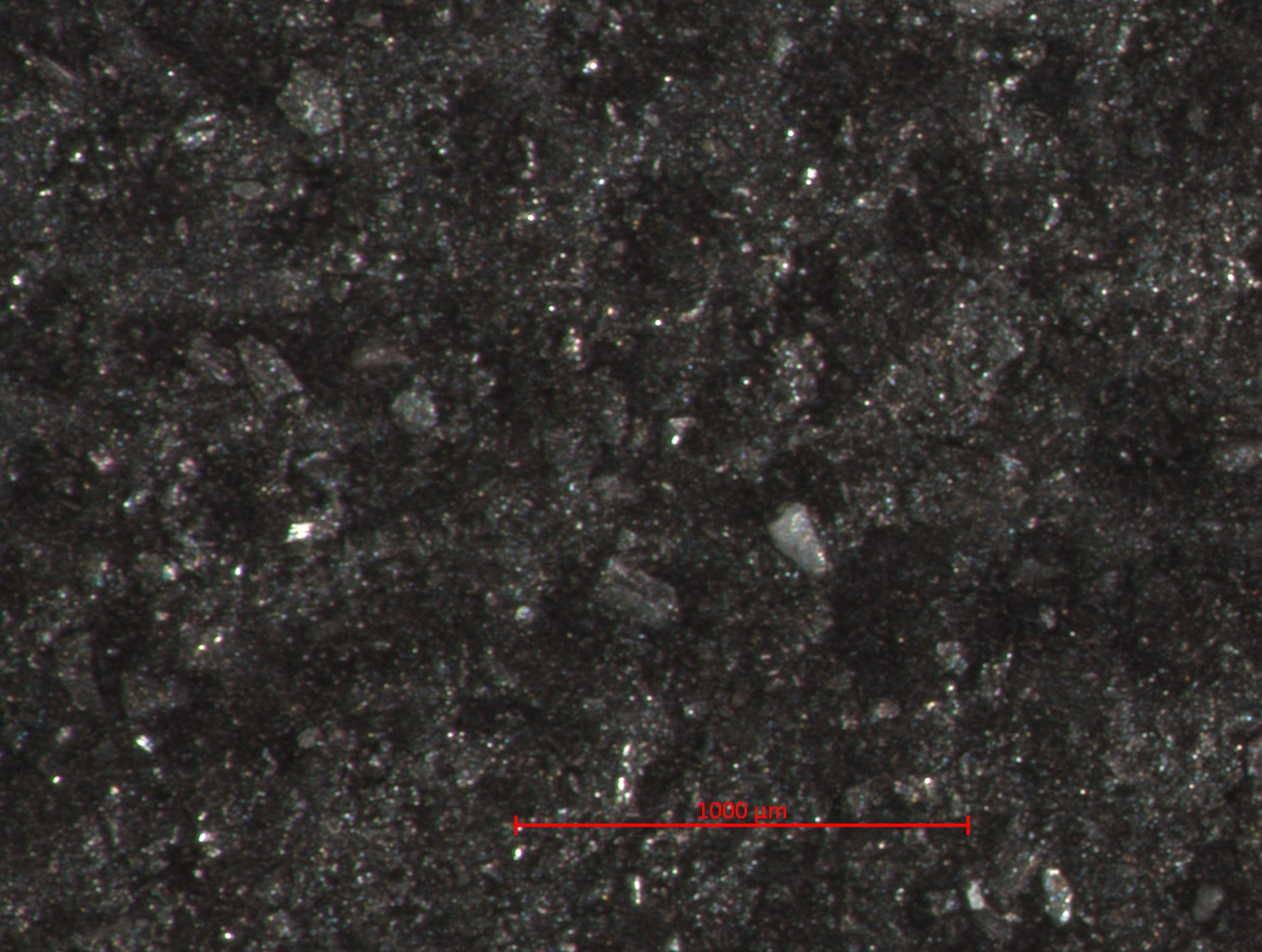
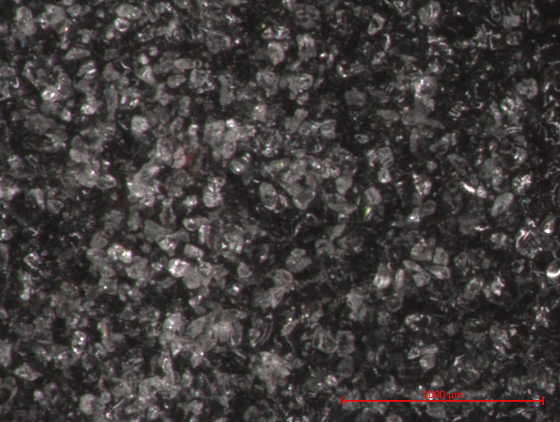
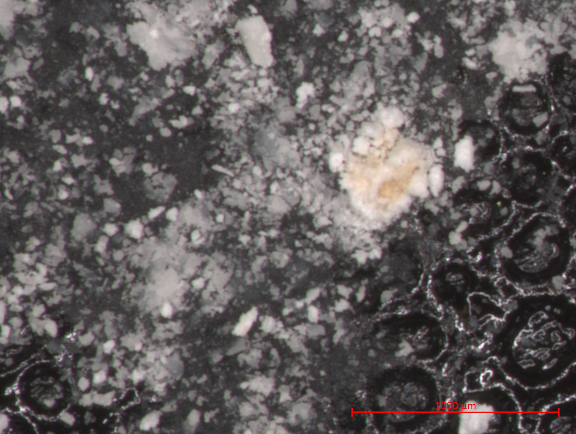
| 15:03, 26 January 2017 | 2 02 6 IvoryBlackWeber STEMI 1000um.jpg (file) |  |
628 KB | Photomicrograph with 1000µm measurement bar (approximate to 50x magnification) (LC) Image captured using Zeiss STEMI SV 11 stereomicroscope. Pigment sample was applied to carbon tape and mounted onto aluminum examination stub. Image was illuminated b... | 1 |
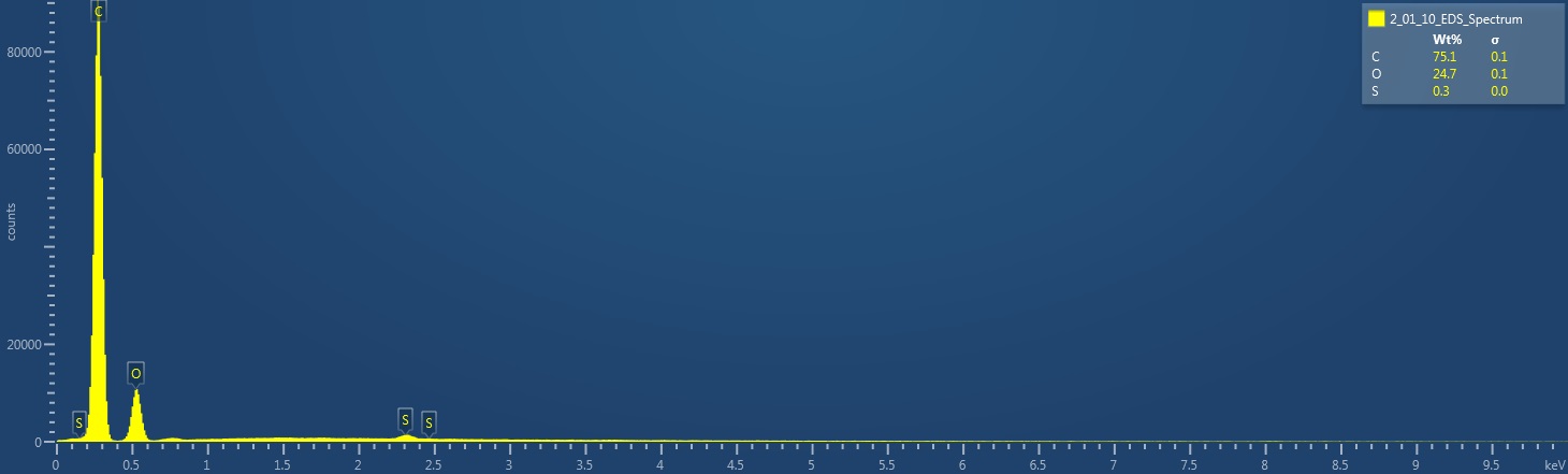
| 15:38, 25 January 2017 | 2 01 10 LampBlack EDS Spectrum.jpg (file) |  |
53 KB | EDS Spectrum showing elemental peak height as a function of X-ray counts collected (LC) Elements Identified Major (> 10%): carbon, oxygen. Minor (1-10%): NA. Trace (< 1%): sulfur. Area X-ray counts collected: 1,012,732. Live Time: 12.9 seconds. Mag... | 1 |
| 15:35, 25 January 2017 | 2 01 10 LampBlack SEM 50um.jpg (file) |  |
499 KB | SEM image with 50µm measurement bar (approximate to 1000x magnification) (LC) Imaged with FEI Quanta 600 scanning electron microscope (SEM) and xT microscope Server user interface. Pigment sample was applied to carbon tape and mounted onto aluminum e... | 1 |
| 15:32, 25 January 2017 | 2 01 10 LampBlack SEM 100um.jpg (file) |  |
515 KB | SEM image with 100µm measurement bar (approximate to 500x magnification) (LC) Imaged with FEI Quanta 600 scanning electron microscope (SEM) and xT microscope Server user interface. Pigment sample was applied to carbon tape and mounted onto aluminum e... | 1 |
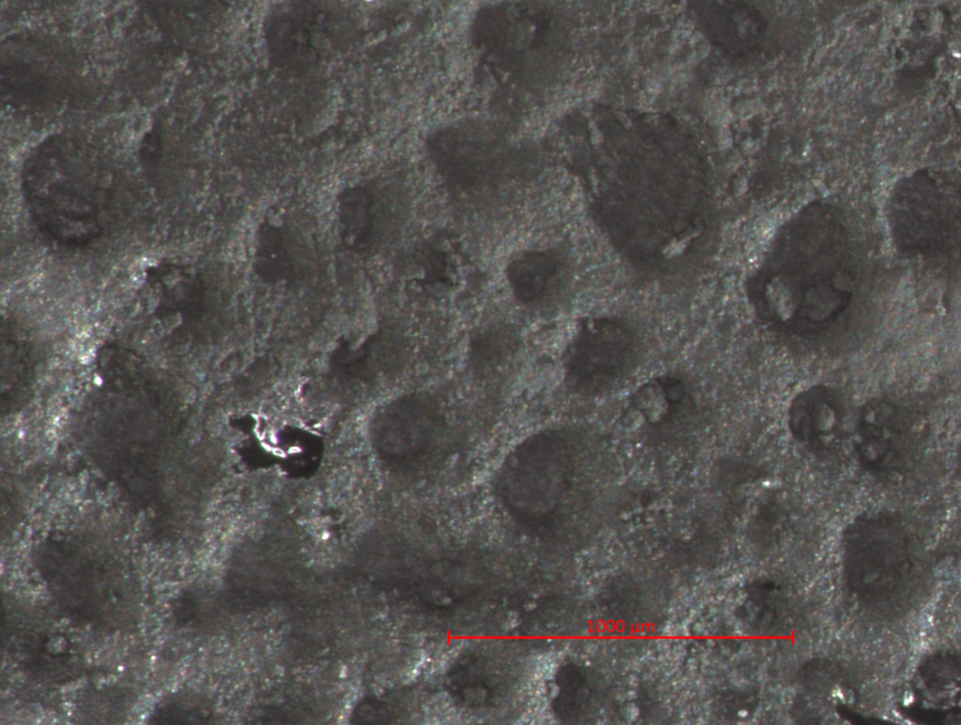
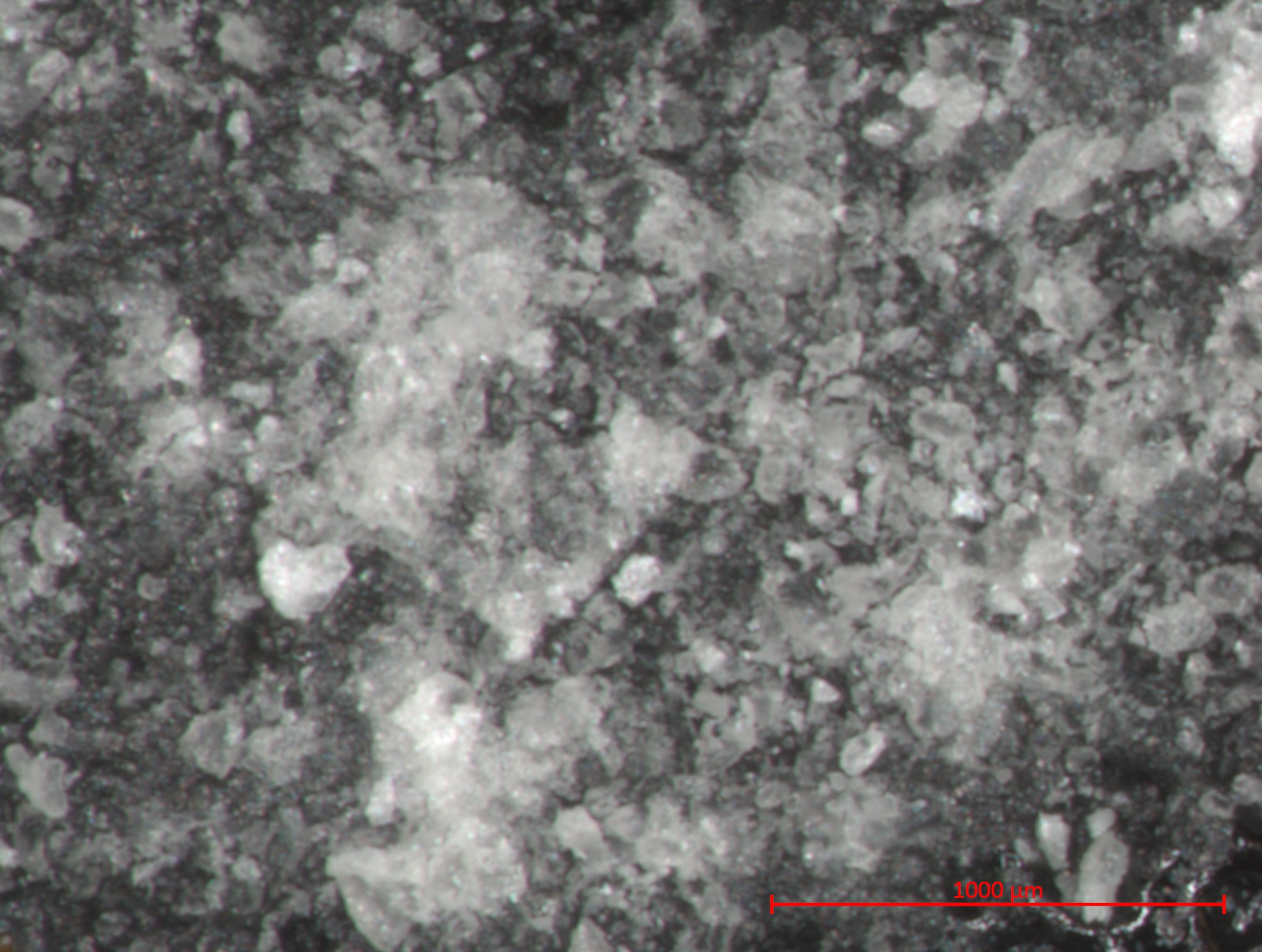
| 15:31, 25 January 2017 | 2 01 10 LampBlack STEMI 1000um.jpg (file) |  |
652 KB | Photomicrograph with 1000µm measurement bar (approximate to 50x magnification) (LC) Image captured using Zeiss STEMI SV 11 stereomicroscope. Pigment sample was applied to carbon tape and mounted onto aluminum examination stub. Image was illuminated b... | 1 |
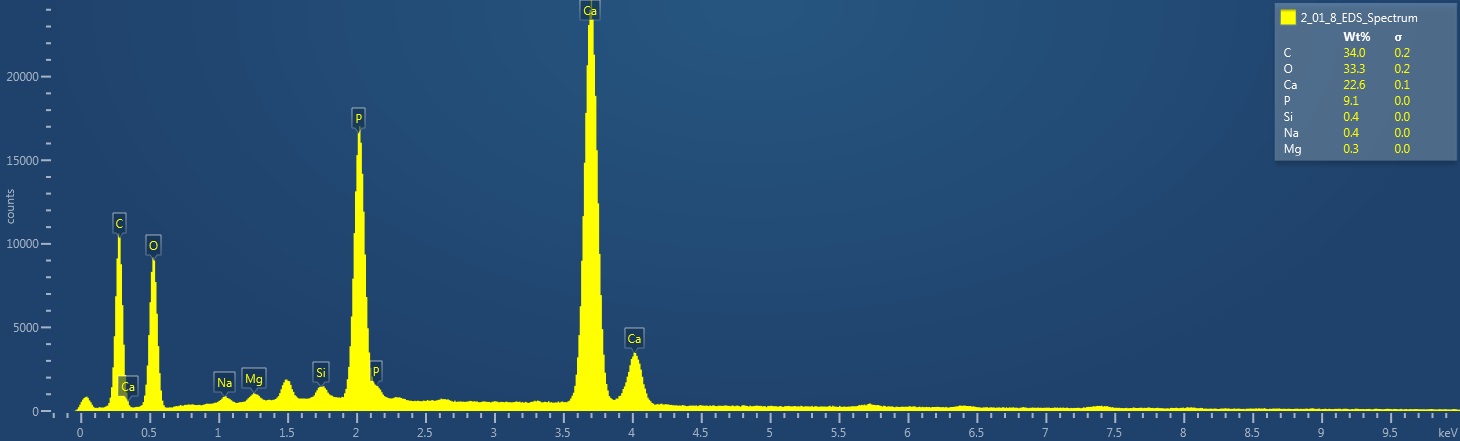
| 15:11, 25 January 2017 | 2 01 8 LampblackTrimount EDS Spectrum.jpg (file) |  |
70 KB | EDS Spectrum showing elemental peak height as a function of X-ray counts collected (LC) Elements Identified Major (> 10%): carbon, oxygen, calcium. Minor (1-10%): phosphorus. Trace (< 1%): silicone, sodium, magnesium. Area X-ray counts collected: 1... | 1 |
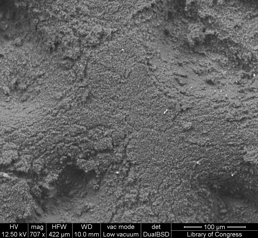
| 15:08, 25 January 2017 | 2 01 8 LampblackTrimount SEM 50um.jpg (file) |  |
489 KB | SEM image with 50µm measurement bar (approximate to 1000x magnification) (LC) Imaged with FEI Quanta 600 scanning electron microscope (SEM) and xT microscope Server user interface. Pigment sample was applied to carbon tape and mounted onto aluminum e... | 1 |
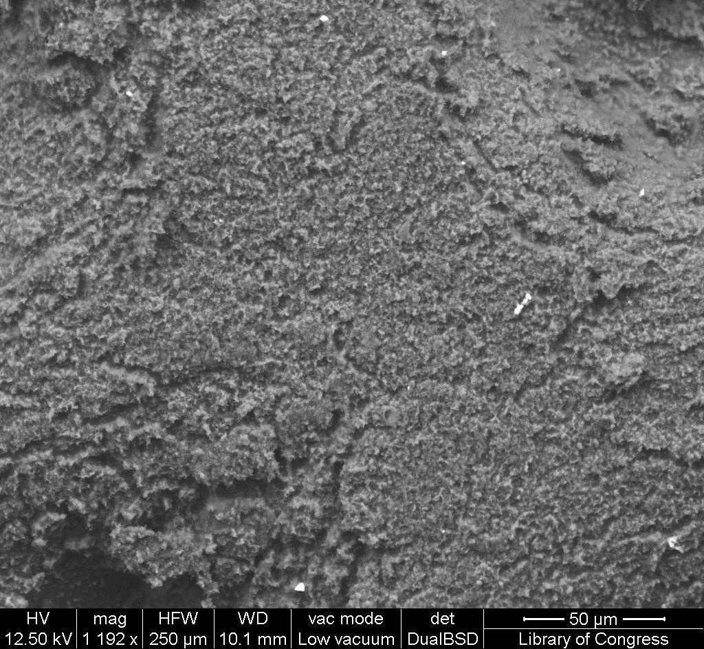
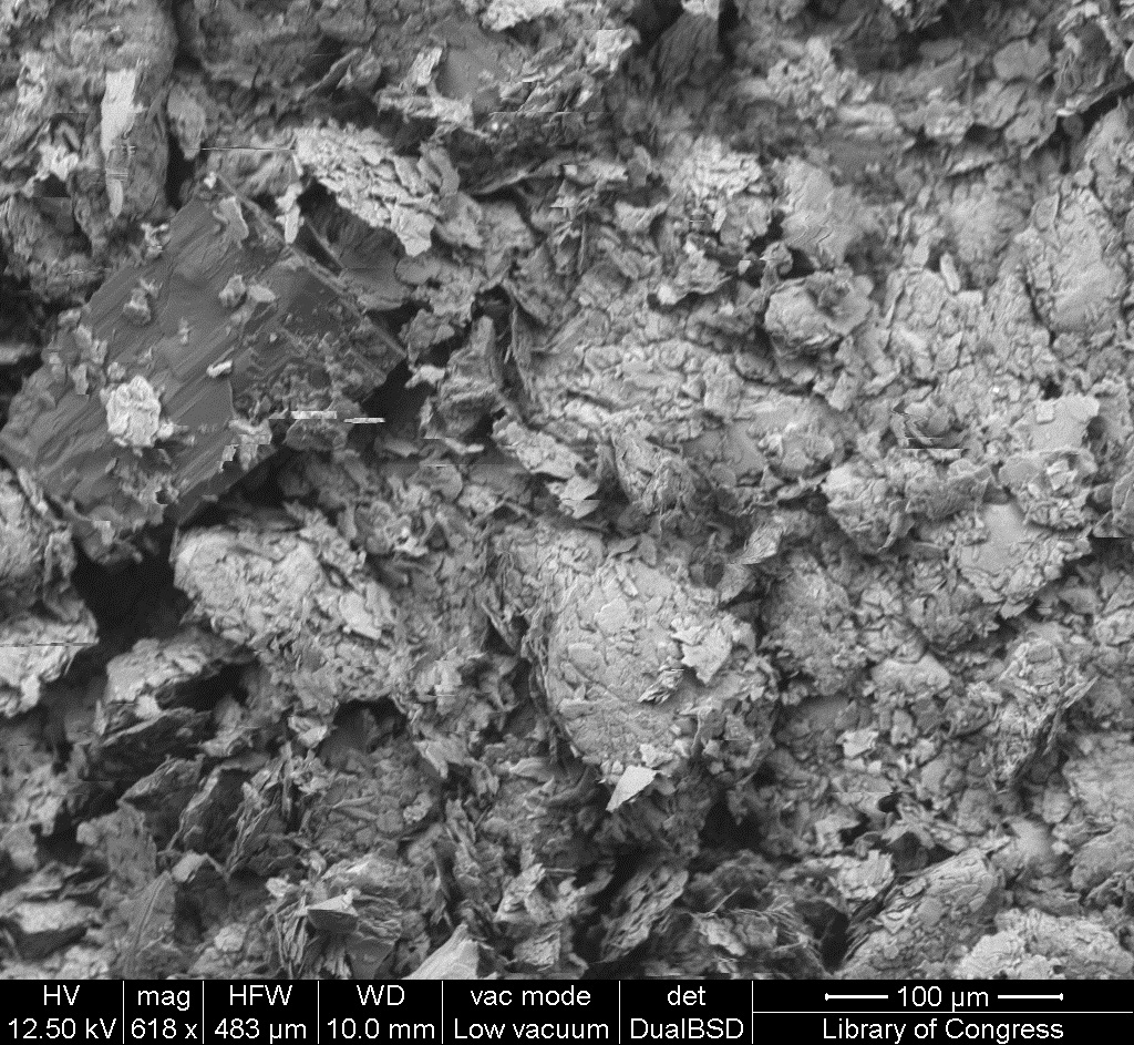
| 15:07, 25 January 2017 | 2 01 8 LampblackTrimount SEM 100um.jpg (file) |  |
546 KB | SEM image with 100µm measurement bar (approximate to 500x magnification) (LC) Imaged with FEI Quanta 600 scanning electron microscope (SEM) and xT microscope Server user interface. Pigment sample was applied to carbon tape and mounted onto aluminum e... | 1 |
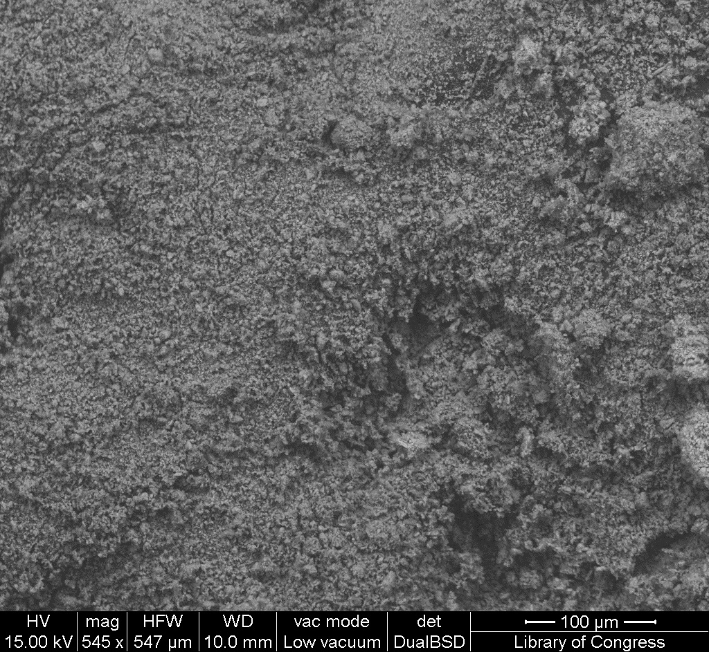
| 15:06, 25 January 2017 | 2 01 8 LampblackTrimount STEMI 1000um.jpg (file) |  |
600 KB | Photomicrograph with 1000µm measurement bar (approximate to 50x magnification) (LC) Image captured using Zeiss STEMI SV 11 stereomicroscope. Pigment sample was applied to carbon tape and mounted onto aluminum examination stub. Image was illuminated b... | 1 |
| 14:20, 25 January 2017 | 2 01 7 Charcoal EDS Spectrum.jpg (file) |  |
59 KB | EDS Spectrum showing elemental peak height as a function of X-ray counts collected (LC) Elements Identified Major (> 10%): carbon, oxygen. Minor (1-10%): calcium. Trace (< 1%): iron, silicon, potassium, sulfur. Area X-ray counts collected: 1,015,88... | 1 |
| 14:18, 25 January 2017 | 2 01 7 Charcoal SEM 50um.jpg (file) |  |
450 KB | SEM image with 50µm measurement bar (approximate to 1000x magnification) (LC) Imaged with FEI Quanta 600 scanning electron microscope (SEM) and xT microscope Server user interface. Pigment sample was applied to carbon tape and mounted onto aluminum e... | 1 |
| 14:17, 25 January 2017 | 2 01 7 Charcoal SEM 100um.jpg (file) |  |
531 KB | SEM image with 100µm measurement bar (approximate to 500x magnification) (LC) Imaged with FEI Quanta 600 scanning electron microscope (SEM) and xT microscope Server user interface. Pigment sample was applied to carbon tape and mounted onto aluminum e... | 1 |
| 14:16, 25 January 2017 | 2 01 7 Charcoal STEMI 1000um.jpg (file) |  |
626 KB | Photomicrograph with 1000µm measurement bar (approximate to 50x magnification) (LC) Image captured using Zeiss STEMI SV 11 stereomicroscope. Pigment sample was applied to carbon tape and mounted onto aluminum examination stub. Image was illuminated b... | 1 |
| 13:52, 25 January 2017 | 2 01 6 Tierra Negra EDS Spectrum.jpg (file) | 82 KB | EDS Spectrum showing elemental peak height as a function of X-ray counts collected (LC) Elements Identified Major (> 10%): carbon, oxygen, silicon. Minor (1-10%): aluminum, iron, potassium. Trace (< 1%): calcium, magnesium, phosphorus, titanium, sul... | 1 | |
| 13:43, 25 January 2017 | 2 01 6 TierraNegra SEM 50um.jpg (file) |  |
525 KB | SEM image with 50µm measurement bar (approximate to 1000x magnification) (LC) Imaged with FEI Quanta 600 scanning electron microscope (SEM) and xT microscope Server user interface. Pigment sample was applied to carbon tape and mounted onto aluminum e... | 1 |
| 13:42, 25 January 2017 | 2 01 6 TierraNegra SEM 100um.jpg (file) |  |
548 KB | SEM image with 100µm measurement bar (approximate to 500x magnification) (LC) Imaged with FEI Quanta 600 scanning electron microscope (SEM) and xT microscope Server user interface. Pigment sample was applied to carbon tape and mounted onto aluminum e... | 1 |
| 13:41, 25 January 2017 | 2 01 6 TierraNegra STEMI 1000um.jpg (file) |  |
575 KB | Photomicrograph with 1000µm measurement bar (approximate to 50x magnification) (LC) Image captured using Zeiss STEMI SV 11 stereomicroscope. Pigment sample was applied to carbon tape and mounted onto aluminum examination stub. Image was illuminated b... | 1 |
| 14:03, 17 January 2017 | 1 20 7 GofunOyster31 EDS Spectrum.jpg (file) | 72 KB | EDS Spectrum showing elemental peak height as a function of X-ray counts collected (LC) Elements Identified Major (> 10%): oxygen, carbon, calcium. Minor (1-10%): NA. Trace (< 1%): sodium, sulfur, silicon. Area X-ray counts collected: 1,022,313. Li... | 1 | |
| 14:02, 17 January 2017 | 1 20 7 GofunOyster31 SEM 50um.jpg (file) |  |
535 KB | SEM image with 50µm measurement bar (approximate to 1000x magnification) (LC) Imaged with FEI Quanta 600 scanning electron microscope (SEM) and xT microscope Server user interface. Pigment sample was applied to carbon tape and mounted onto aluminum e... | 1 |
| 14:01, 17 January 2017 | 1 20 7 GofunOyster31 SEM 100um.jpg (file) |  |
582 KB | SEM image with 100µm measurement bar (approximate to 500x magnification) (LC) Imaged with FEI Quanta 600 scanning electron microscope (SEM) and xT microscope Server user interface. Pigment sample was applied to carbon tape and mounted onto aluminum e... | 1 |
| 14:00, 17 January 2017 | 1 20 7 GofunOyster31 STEMI 1000um.jpg (file) |  |
495 KB | Photomicrograph with 1000µm measurement bar (approximate to 50x magnification) (LC) Image captured using Zeiss STEMI SV 11 stereomicroscope. Pigment sample was applied to carbon tape and mounted onto aluminum examination stub. Image was illuminated b... | 1 |
| 13:44, 17 January 2017 | 1 20 6 Suisho EDS Spectrum.jpg (file) | 59 KB | EDS Spectrum showing elemental peak height as a function of X-ray counts collected (LC) Elements Identified Major (> 10%): oxygen, silicon, carbon. Minor (1-10%): NA. Trace (< 1%): NA. Area X-ray counts collected: 1,019,126. Live Time: 47.5 seconds... | 1 | |
| 13:42, 17 January 2017 | 1 20 6 Suisho SEM 50um.jpg (file) |  |
411 KB | SEM image with 50µm measurement bar (approximate to 1000x magnification) (LC) Imaged with FEI Quanta 600 scanning electron microscope (SEM) and xT microscope Server user interface. Pigment sample was applied to carbon tape and mounted onto aluminum e... | 1 |
| 13:41, 17 January 2017 | 1 20 6 Suisho SEM 100um.jpg (file) |  |
432 KB | SEM image with 100µm measurement bar (approximate to 500x magnification) (LC) Imaged with FEI Quanta 600 scanning electron microscope (SEM) and xT microscope Server user interface. Pigment sample was applied to carbon tape and mounted onto aluminum e... | 1 |
| 13:40, 17 January 2017 | 1 20 6 Suisho STEMI 1000um.jpg (file) |  |
585 KB | Photomicrograph with 1000µm measurement bar (approximate to 50x magnification) (LC) Image captured using Zeiss STEMI SV 11 stereomicroscope. Pigment sample was applied to carbon tape and mounted onto aluminum examination stub. Image was illuminated b... | 1 |
| 13:18, 17 January 2017 | 1 20 5 Kira42 EDS Spectrum.jpg (file) | 67 KB | EDS Spectrum showing elemental peak height as a function of X-ray counts collected (LC) Elements Identified Major (> 10%): oxygen, carbon, silicon, magnesium. Minor (1-10%): NA. Trace (< 1%): NA. Area X-ray counts collected: 1,022,797. Live Time: 5... | 1 | |
| 13:16, 17 January 2017 | 1 20 5 Kira42 SEM 50um.jpg (file) |  |
476 KB | SEM image with 50µm measurement bar (approximate to 1000x magnification) (LC) Imaged with FEI Quanta 600 scanning electron microscope (SEM) and xT microscope Server user interface. Pigment sample was applied to carbon tape and mounted onto aluminum e... | 1 |
| 13:14, 17 January 2017 | 1 20 5 Kira42 SEM 100um.jpg (file) |  |
498 KB | SEM image with 100µm measurement bar (approximate to 500x magnification) (LC) Imaged with FEI Quanta 600 scanning electron microscope (SEM) and xT microscope Server user interface. Pigment sample was applied to carbon tape and mounted onto aluminum e... | 1 |
| 13:13, 17 January 2017 | 1 20 5 Kira42 STEMI 1000um.jpg (file) |  |
567 KB | Photomicrograph with 1000µm measurement bar (approximate to 50x magnification) (LC) Image captured using Zeiss STEMI SV 11 stereomicroscope. Pigment sample was applied to carbon tape and mounted onto aluminum examination stub. Image was illuminated b... | 1 |
| 11:02, 17 January 2017 | 1 20 4 GofunOysterShell EDS Spectrum.jpg (file) | 69 KB | EDS Spectrum showing elemental peak height as a function of X-ray counts collected (LC) Elements Identified Major (> 10%): oxygen, carbon, calcium. Minor (1-10%): NA. Trace (< 1%): sulfur. Area X-ray counts collected: 1,028,594. Live Time: 66.0 sec... | 1 | |
| 11:01, 17 January 2017 | 1 20 4 GofunOysterShell SEM 50um.jpg (file) |  |
473 KB | SEM image with 50µm measurement bar (approximate to 1000x magnification) (LC) Imaged with FEI Quanta 600 scanning electron microscope (SEM) and xT microscope Server user interface. Pigment sample was applied to carbon tape and mounted onto aluminum e... | 1 |
| 11:00, 17 January 2017 | 1 20 4 GofunOysterShell SEM 100um.jpg (file) |  |
552 KB | SEM image with 100µm measurement bar (approximate to 500x magnification) (LC) Imaged with FEI Quanta 600 scanning electron microscope (SEM) and xT microscope Server user interface. Pigment sample was applied to carbon tape and mounted onto aluminum e... | 1 |
| 10:59, 17 January 2017 | 1 20 4 GofunOysterShell STEMI 1000um.jpg (file) |  |
571 KB | Photomicrograph with 1000µm measurement bar (approximate to 50x magnification) (LC) Image captured using Zeiss STEMI SV 11 stereomicroscope. Pigment sample was applied to carbon tape and mounted onto aluminum examination stub. Image was illuminated b... | 1 |
| 10:50, 17 January 2017 | 1 20 3 GofunSeaShell EDS Spectrum.jpg (file) | 69 KB | EDS Spectrum showing elemental peak height as a function of X-ray counts collected (LC) Elements Identified Major (> 10%): oxygen, carbon, calcium. Minor (1-10%): NA. Trace (< 1%): sulfur. Area X-ray counts collected: 1,029,212. Live Time: 70.5 sec... | 1 | |
| 10:49, 17 January 2017 | 1 20 3 GofunSeaShell SEM 50um.jpg (file) |  |
497 KB | SEM image with 50µm measurement bar (approximate to 1000x magnification) (LC) Imaged with FEI Quanta 600 scanning electron microscope (SEM) and xT microscope Server user interface. Pigment sample was applied to carbon tape and mounted onto aluminum e... | 1 |
| 10:48, 17 January 2017 | 1 20 3 GofunSeaShell SEM 100um.jpg (file) |  |
528 KB | SEM image with 100µm measurement bar (approximate to 500x magnification) (LC) Imaged with FEI Quanta 600 scanning electron microscope (SEM) and xT microscope Server user interface. Pigment sample was applied to carbon tape and mounted onto aluminum e... | 1 |
| 10:47, 17 January 2017 | 1 20 3 GofunSeaShell STEMI 1000um.jpg (file) |  |
506 KB | Photomicrograph with 1000µm measurement bar (approximate to 50x magnification) (LC) Image captured using Zeiss STEMI SV 11 stereomicroscope. Pigment sample was applied to carbon tape and mounted onto aluminum examination stub. Image was illuminated b... | 1 |
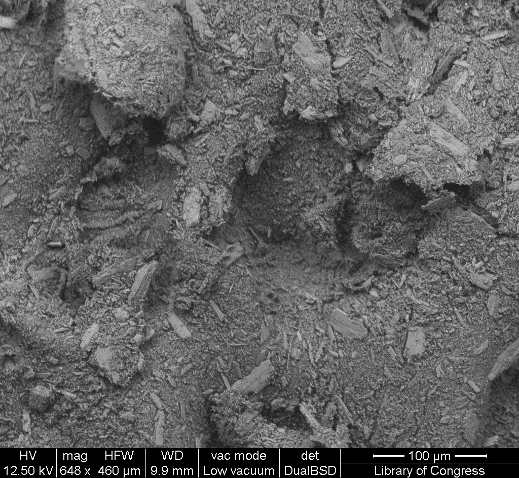
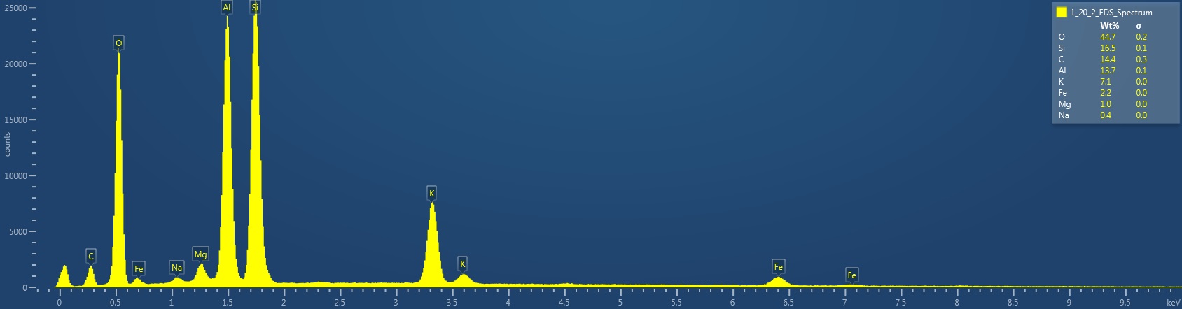
| 15:08, 12 January 2017 | 1 20 2 Kiara EDS Spectrum.jpg (file) | 78 KB | EDS Spectrum showing elemental peak height as a function of X-ray counts collected (LC) Elements Identified Major (> 10%): oxygen, silicon, carbon, aluminum. Minor (1-10%): potassium, iron, magnesium. Trace (< 1%): sodium. Area X-ray counts collect... | 1 | |
| 15:05, 12 January 2017 | 1 20 2 Kiara SEM 50um.jpg (file) |  |
416 KB | SEM image with 50µm measurement bar (approximate to 1000x magnification) (LC) Imaged with FEI Quanta 600 scanning electron microscope (SEM) and xT microscope Server user interface. Pigment sample was applied to carbon tape and mounted onto aluminum e... | 1 |
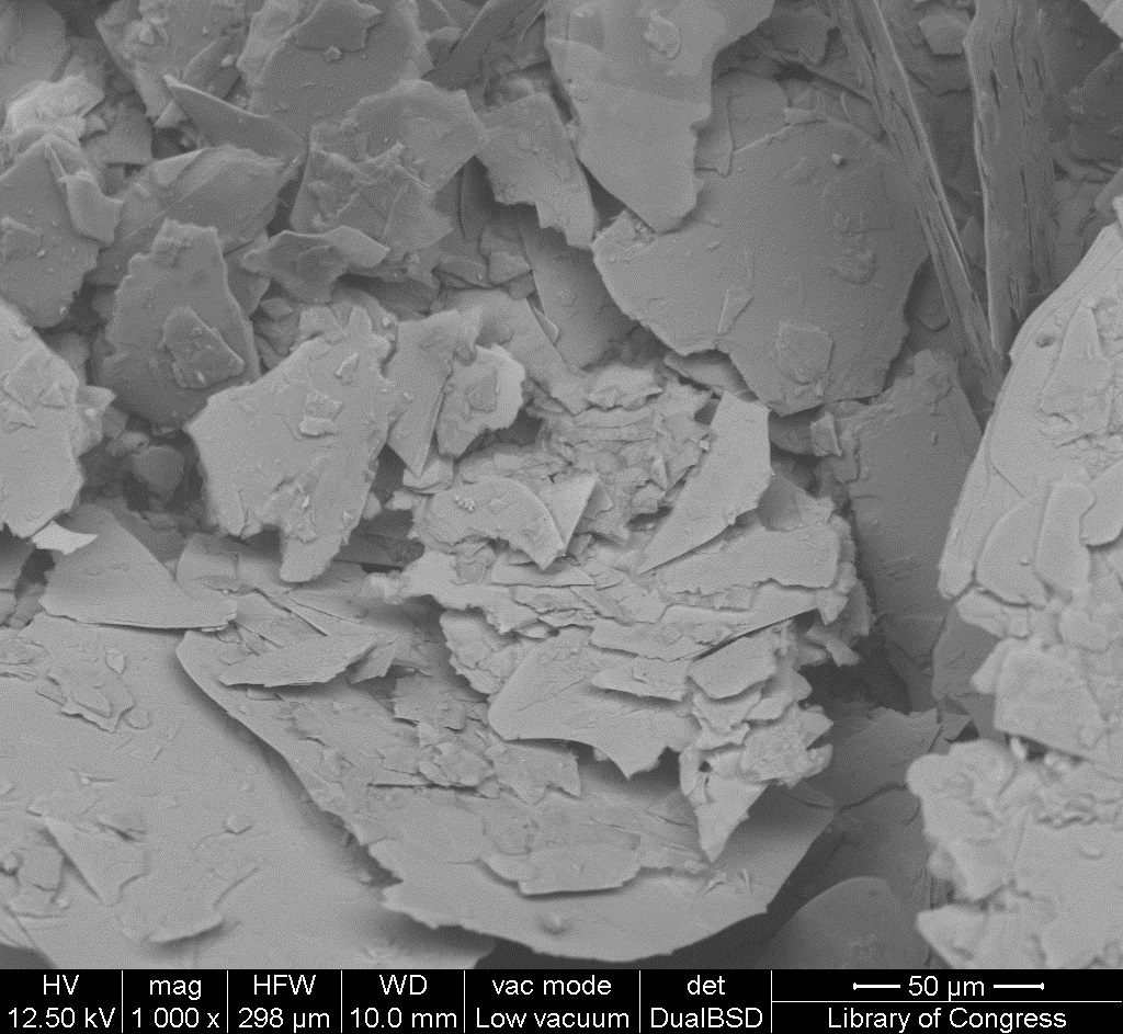
| 15:05, 12 January 2017 | 1 20 2 Kiara SEM 100um.jpg (file) |  |
442 KB | SEM image with 100µm measurement bar (approximate to 500x magnification) (LC) Imaged with FEI Quanta 600 scanning electron microscope (SEM) and xT microscope Server user interface. Pigment sample was applied to carbon tape and mounted onto aluminum e... | 1 |
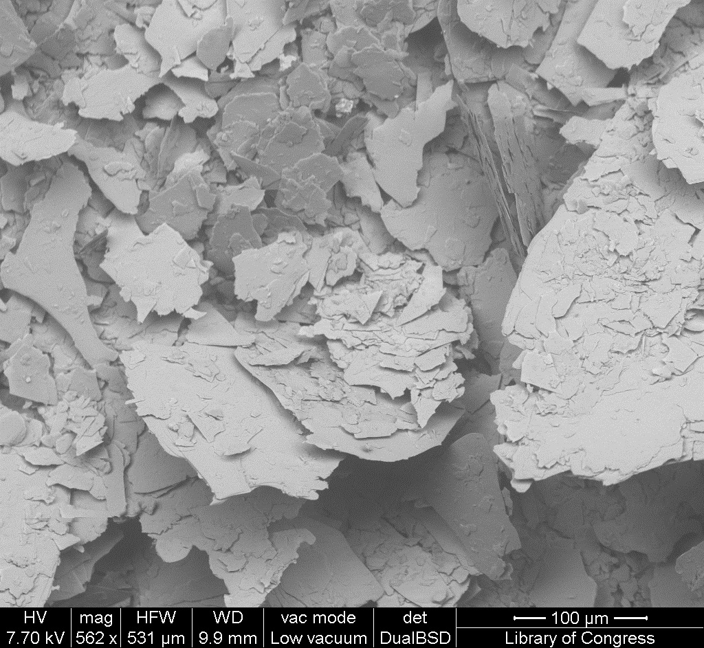
| 15:03, 12 January 2017 | 1 20 2 Kiara STEMI 1000um.jpg (file) |  |
524 KB | Photomicrograph with 1000µm measurement bar (approximate to 50x magnification) (LC) Image captured using Zeiss STEMI SV 11 stereomicroscope. Pigment sample was applied to carbon tape and mounted onto aluminum examination stub. Image was illuminated b... | 1 |
| 14:20, 12 January 2017 | 1 10 3 NoLabel EDS Spectrum.jpg (file) | 68 KB | EDS Spectrum showing elemental peak height as a function of X-ray counts collected (LC) Elements Identified Major (> 10%): oxygen, carbon, silicon, magnesium. Minor (1-10%): NA. Trace (< 1%): NA. Area X-ray counts collected: 1,047,913. Live Time: 4... | 1 | |
| 14:17, 12 January 2017 | 1 10 3 NoLabel SEM 50um.jpg (file) |  |
493 KB | SEM image with 50µm measurement bar (approximate to 1000x magnification) (LC) Imaged with FEI Quanta 600 scanning electron microscope (SEM) and xT microscope Server user interface. Pigment sample was applied to carbon tape and mounted onto aluminum e... | 1 |
| 14:17, 12 January 2017 | 1 10 3 NoLabel SEM 100um.jpg (file) |  |
527 KB | SEM image with 100µm measurement bar (approximate to 500x magnification) (LC) Imaged with FEI Quanta 600 scanning electron microscope (SEM) and xT microscope Server user interface. Pigment sample was applied to carbon tape and mounted onto aluminum e... | 1 |
| 14:16, 12 January 2017 | 1 10 3 NoLabel STEMI 1000um.jpg (file) |  |
594 KB | Photomicrograph with 1000µm measurement bar (approximate to 50x magnification) (LC) Image captured using Zeiss STEMI SV 11 stereomicroscope. Pigment sample was applied to carbon tape and mounted onto aluminum examination stub. Image was illuminated b... | 1 |
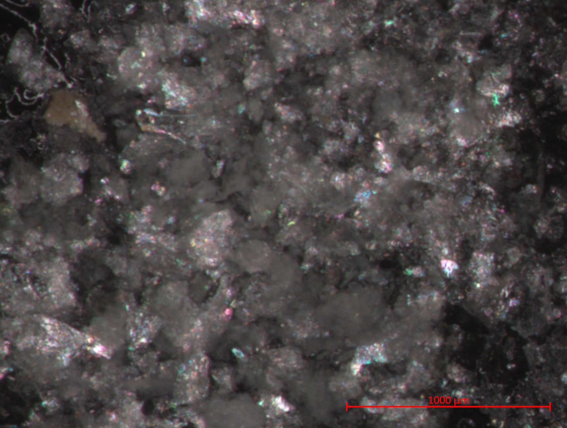
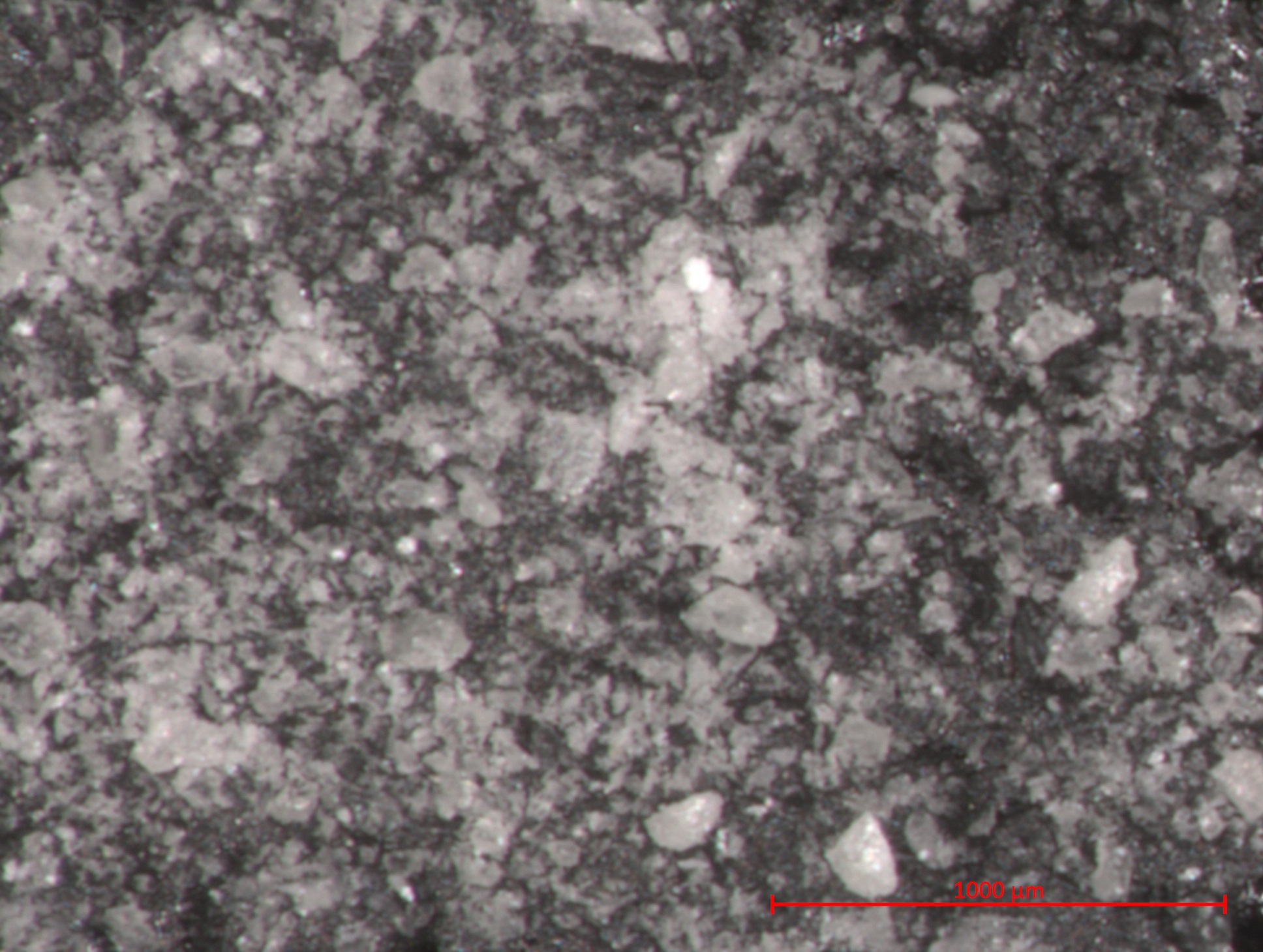
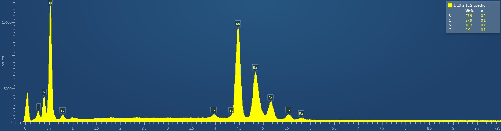
| 13:54, 12 January 2017 | 1 10 2 WhiteSpain EDS Spectrum.jpg (file) | 78 KB | EDS Spectrum showing elemental peak height as a function of X-ray counts collected (LC) Elements Identified Major (> 10%): barium, oxygen, nitrogen. Minor (1-10%): carbon. Trace (< 1%): NA. Area X-ray counts collected: 1,036,309. Live Time: 90.7 se... | 1 |