Search results
Jump to navigation
Jump to search
EV 106 VIS 100x annotated.jpg ...roscope with EXFO X-cite 120 fluorescence illumination system with halogen light source. Spot Flex digital camera with Spot Advance v.4.6 software. Photomic(720 × 720 (470 KB)) - 09:21, 20 May 2015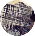
Microcline.jpg Polarizing microscope picture of thin section taken in crossed polarized light showing characteristic twinning cross-hatching(500 × 510 (181 KB)) - 13:10, 19 December 2022
Yellow XPL 1000x.JPG Nikon NiU polarizing light microscope, Nikon DSRi1 camera, Nikon BR Elements software.(844 × 674 (52 KB)) - 11:04, 30 December 2020
Yellow PPL 1000x.JPG Nikon NiU polarizing light microscope, Nikon DSRi1 camera, Nikon BR Elements software.(848 × 675 (98 KB)) - 11:04, 30 December 2020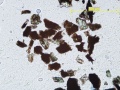
ChartImageLarge 2369.jpg Micrograph, normal light (ICA) ...ing and fractured structures. Twinning was also seen under cross polarized light. Most of the pigment particles are blackened, and exhibit the evidence of h(638 × 480 (67 KB)) - 10:33, 4 May 2013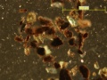
ChartImageLarge 2370.jpg Micrograph, reflected light (ICA) ...ing and fractured structures. Twinning was also seen under cross polarized light. Most of the pigment particles are blackened, and exhibit the evidence of h(638 × 480 (44 KB)) - 10:33, 4 May 2013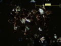
ChartImageLarge 2371.jpg Micrograph, polarized light (ICA) ...ing and fractured structures. Twinning was also seen under cross polarized light. Most of the pigment particles are blackened, and exhibit the evidence of h(638 × 480 (30 KB)) - 10:33, 4 May 2013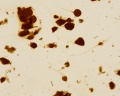
Asphaltum PPL 200x.jpg ...phaltum in PPL at 200X. Examined with a Nikon NiU upright polarizing light microscope, image captured with DSRi1 Pixel-shifting camera and Nikon Elements Basic R(1,236 × 989 (842 KB)) - 14:37, 27 September 2017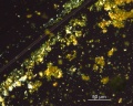
Orpiment XPL 400x.jpg ...r)in PPL at 400X. Image captured with a Nikon NiU upright polarizing light microscope and DSRi1 Pixel-shifting camera + Nikon Elements Basic Research software. C(875 × 700 (646 KB)) - 09:06, 4 October 2017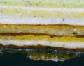
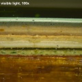
Reid House RE 38 200x.JPG Sample RE 38, visible light, 200x. Nikon NiU epi-fluorescence microscope with Nikon DSRi1 camera, Nikon BR Elements software.(844 × 670 (83 KB)) - 12:19, 30 December 2020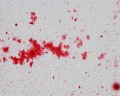
Alizarin Crimson PPL 400x.jpg ...Crimson in PPL at 400X. Examined with a Nikon NiU upright polarizing light microscope, image captured with DSRi1 Pixel-shifting camera and Nikon Elements Basic R(1,168 × 934 (925 KB)) - 13:39, 27 September 2017
Asphaltum XPL 200x.jpg ...Material is isotropic. Examined with a Nikon NiU upright polarizing light microscope, image captured with DSRi1 Pixel-shifting camera and Nikon Elements Basic R(1,236 × 989 (728 KB)) - 14:37, 27 September 2017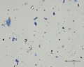
Lapis Natural PPL 400x.JPG ...(Kremer)in PPL at 400X. Examined with a Nikon NiU upright polarizing light microscope, image captured with DSRi1 Pixel-shifting camera and Nikon Elements Basic R(999 × 798 (89 KB)) - 14:13, 29 September 2017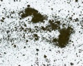
19thc Naples Yellow lead zinc antimonate Kremer PPL2 400x.jpg ...(Kremer)in PPL at 400X. Examined with a Nikon NiU upright polarizing light microscope, image captured with DSRi1 Pixel-shifting camera and Nikon Elements Basic R(1,168 × 934 (963 KB)) - 13:31, 27 September 2017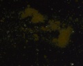
19thc Naples Yellow lead zinc antimonate Kremer XPL2 400x.jpg ...(Kremer)in XPL at 400X. Examined with a Nikon NiU upright polarizing light microscope, image captured with DSRi1 Pixel-shifting camera and Nikon Elements Basic R(1,236 × 989 (783 KB)) - 13:32, 27 September 2017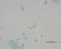
Blue verditer PPL 400x.jpg ...(n=1.662), PPL at 400X. Examined with a Nikon NiU upright polarizing light microscope, image captured with DSRi1 Pixel-shifting camera and Nikon Elements Basic R(1,038 × 830 (637 KB)) - 15:56, 27 September 2017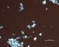
Blue verditer XPL 400x.jpg ...(n=1.662), XPL at 400X. Examined with a Nikon NiU upright polarizing light microscope, image captured with DSRi1 Pixel-shifting camera and Nikon Elements Basic R(1,038 × 830 (662 KB)) - 15:57, 27 September 2017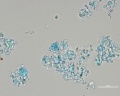
Blue bice verditer PPL 1000x.jpg ...n=1.662), PPL at 1000X. Examined with a Nikon NiU upright polarizing light microscope, image captured with DSRi1 Pixel-shifting camera and Nikon Elements Basic R(1,038 × 830 (696 KB)) - 15:57, 27 September 2017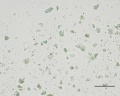
Green verditer Synthesized malachite PPL 400x.jpg ...(n=1.662), PPL at 400X. Examined with a Nikon NiU upright polarizing light microscope, image captured with DSRi1 Pixel-shifting camera and Nikon Elements Basic R(1,038 × 830 (647 KB)) - 15:59, 27 September 2017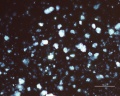
Green verditer Synthesized malachite XPL 400x.jpg ...(n=1.662), XPL at 400X. Examined with a Nikon NiU upright polarizing light microscope, image captured with DSRi1 Pixel-shifting camera and Nikon Elements Basic R(1,038 × 830 (761 KB)) - 15:59, 27 September 2017


