Derrick Fluorescence reds max.jpg

Original file (960 × 720 pixels, file size: 81 KB, MIME type: image/jpeg)
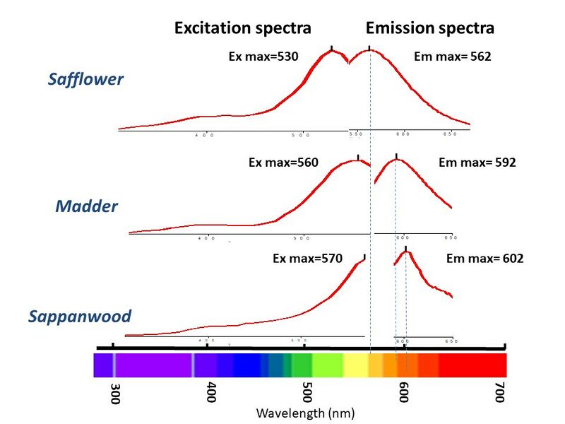
Excitation can occur at many wavelengths. When we set the instrument to scan through several excitation wavelengths, we can determine which wavelength excites the most molecules to fluoresce. This is called the Excitation maxima. (safflower, madder, sappanwood) Exposing the compound to this maximum excitation wavelength we can measure the emission curve And the point with the highest emission is called the Emission max. (safflower, madder, sappanwood) As you can see for these three reds, their Excitation and Emission maxima are distinctly different. Thus these three reds can be distinguished by their fluorescence measurements. Also we can see from these maxima that the emission for safflower is in the yellow region, madder is in the orange region and sappanwood is in the red region.
Our EEM instrument is set to step through the excitation wavelengths every 10 nm and collects a full emission curve at each excitation wavelength. So Instead of looking at the single line plots, we usually look at the contour curve.
File history
Click on a date/time to view the file as it appeared at that time.
| Date/Time | Thumbnail | Dimensions | User | Comment | |
|---|---|---|---|---|---|
| current | 12:41, 22 June 2023 | | 960 × 720 (81 KB) | MDerrick (talk | contribs) |
You cannot overwrite this file.
File usage
The following 2 pages use this file: